- Submissions

Full Text
Novel Research in Sciences
In Vitro Propagation of Juniperus Phoenicea and Juniperus Thurifera from Adult Plant Explants
Ivan Pace and Paola Maria Chiavazza*
Department of Agronomy, Forest and Land Management, Faculty of Agriculture, Italy
*Corresponding author: Ivan Pace, Paola Maria Chiavazza, Department of Agronomy, Forest and Land Management, Faculty of Agriculture, University of Turin, 10095, Turin, Italy
Submission: December 17, 2022;Published: January 21, 2022
.jpg)
Volume10 Issue3January, 2022
Introduction
Studies concerning Juniperus phoenicea L. and Juniperus thurifera L. are aimed at verifying the effectiveness of propagation techniques as alternative conservation strategies on rare spontaneous plant species [1]. The two species are very localized at regional level and therefore have a high conservation interest [2]. J. phoenicea is a species belonging to the Cupressaceae family and is native to some regions of the Mediterranean basin, the Canary Islands and North Africa. J. thurifera is an endemic species that lives in semi-arid mountain environments and grows exclusively in Western Mediterranean (France, Spain, Italy, Morocco and Algeria) (Figure 1).
Figure 1: J. thurifera in Rocca San giovanni-saben Nature Reserve (CN).
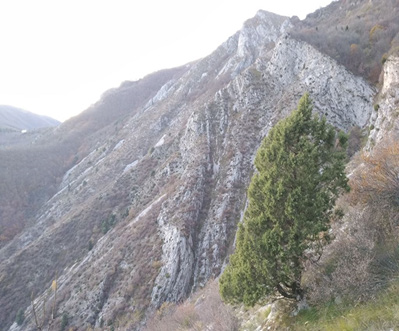
The largest population of J. phoenicea in the Ligurian and Maritime Alps, is present in the special nature reserve “Population of Juniperus phoenicea of Rocca S. Giovanni-Saben” and is considered the highest and northernmost settlement of the species in Italy. J. phoenicea does not propagate effectively with traditional cutting methods [3] and produces a minimal amount of viable seeds. Also, in J. thurifera, the scarcity of viable seeds limits germination. The age of the specimens presents in the stand, excessive grazing, forest cuts, fires and the lack of planned management beyond are limiting abiotic factors [4].
Materials and Methods
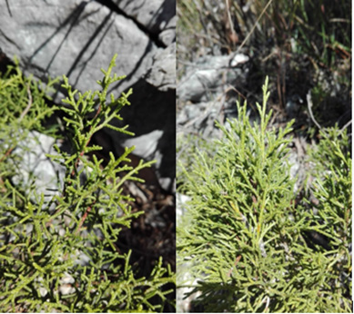
Branches of mother plants of both species (Figure 2) were sampled in nature, in Rocca San Giovanni-Saben Nature Reserve (CN). Collected in the autumn, the plants were used as source of microcuttings for in vitro studies. The twigs were moistened and stored for a few days in a plastic bag at 4 °C in the refrigerator. No preconditioned in a greenhouse was done. The preconditioning consists in the maintenance of explants at 22 °C, 16h photoperiod for at least one week with periodic sprays of a fungicide solution: 0.75g/l Derosal and 1.5g/l Previcur (Hoescht and Schering AgrEvo, Berlin, Germany).
Figure 2: J. phoenicea (sx) and J. thurifera (dx).
Next culture media were respectively used, with plant growth regulators:
1. DKW (Driver and Kuniyaki Walnut)+ BAP 0.1mg/l for J. phoenicea
2. WPM (Woody Plant Medium) + BAP 0.5mg/l +2.4D 0.25mg/l + activated charcoal for J. thurifera.
Sterilization and avoiding of leaf browning
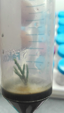
The sterilization operations greatly influenced the study as both species, perhaps due to their morphological characteristics, are very subject to fungal pollution (Figure 3) and leaf browning. NaOCl was used at different concentrations and times, alone or in combination with alcohol, without positive results.
Figure 3: Fungi growth in Juniperus thurifera.
1. 30’ NaOCl 1% + Tween 20
2. 60’ NaOCl 0.5% + Tween 20
3. 1’ alcohol 70% v/v + 30’ NaOCl 1% + Tween 20
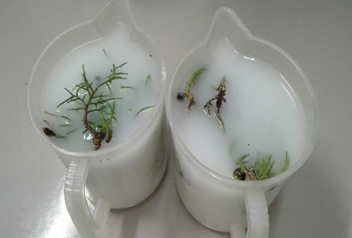
After several negative results, to counteract persistent fungal pollution, NaOCl was associated with a fungicide (Enovit Metil, Figure 4).
Figure 4: Treatment with fungicide on both species.
1. 30’ fungicide (1ml/l) + 15’ NaOCl 1% + Tween 20
2. 30’ fungicide (1ml/l) + 30’ NaOCl 1% + Tween 20
3. 2h fungicide (1ml/l) + 5’ NaOCl 2.5% + Tween 20
To contain the inhibitory action of the endogenous auxins during the formation of new buds (dominance elimination), small explants were placed horizontally (Figure 5). Substrate was first constituted only by agar, leading to the formation of small amounts of callus in J. thurifera (Figure 6).
Figure 5: Horizontal arrangement of the apexes.
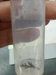
Figure 6: Growth of callus in J. thurifera.
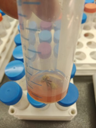
The combinations used were:
1. Quick immersion in fungicide + 1h NaOCl 1% + Tween 20
2. Quick immersion in fungicide + 15’ NaOCl 2.5% + Tween 20
To counteract the still present pollution derived from fungi and widespread leaf browning, the concentration of fungicide was increased (up to 1h fungicide 2ml/l + 1h NaOCl 1% + Tween 20).
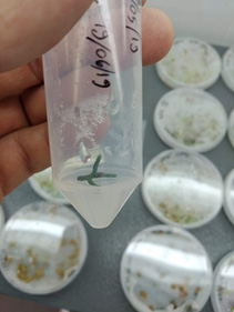
Subsequently, with the add of citric and ascorbic acids (150mg/l) in the NaOCl wash, better results were obtained. Citric and ascorbic acids were added also directly in the culture medium: this action helped to slow down leaf browning (DKW + BAP 0.1mg/l + 150mg/l citric and ascorbic acids for J. phoenicea) and WPM + BAP 0.5mg/l + 2,4D 0.25mg/l + 150mg/l citric and ascorbic acids for J. thurifera, (Figure 7).
Figure 7: Antioxidant effect of the culture medium on the apexes.
Nodal explants and callus multiplication
Plant tissues culture of both species was carried out by testing the response at some frequently used culture media.
For J. phoenicea was utilized DKW + BAP 0.1mg/l [5].
For J. thurifera was preferred WPM + BAP 0.5 and 2.4D 0.25mg/l + activated charcoal [4].
To contain the inhibitory action of endogenous auxins in the formation of new buds, some explants of both species were positioned horizontally on the medium, using MS as substrate. After about 30 days, the explants of J. thurifera browned over time and began to produce callus. It has been noted that calluses are also produced by browned explants transferred in a horizontal position, even if previously held in a vertical position. The Calli were left to grow on the explants and once they reached dimensions suitable for their division, they were transferred to a medium for callus culture. The only protocol found in bibliography refers to J. communis, a similar species that shares in part the habitat of both species under study.
The medium chosen for the culture and multiplication of callus was MS + 2,4D 0.5 and BAP 0.1mg/l. Cultures were incubated at (25±2)°C and were transferred to fresh medium every month. It has been noticed that Calli reach a maximum growth size then they darken, probably due to the phenols they produce. The transfer of the still green masses is essential for a good multiplication phase (Figure 8). The latest research is aimed to obtain somatic embryogenesis on J. thurifera callus, transferring part of the available explant stock onto an induction medium consisting of MS + 2.4D 1mg/l. A series of Falcon tubes were prepared and then divided into two stocks: to verify the ideal growth conditions for the calli, half of them were placed at 20 °C light (photoperiod 12/12h) and the other half at 20 °C dark. After one month, part of them will be transferred to a MS determination medium with pH 5.4 for the differentiation of the embryos.
Figure 8: Green callus in J. thurifera.

Result
Due to the many difficulties encountered during sterilization operations, it has been understood that the time of action of the fungicide in association with NaOCl is essential for maintaining the sterility of the explants. The browning, due to the production of phenols by the explants, was effectively counteracted with the use of citric acid and ascorbic acid for J. phoenicea, together with activated carbon for J. thurifera. The production of phenols could be induced by the abundance of salts in the culture medium, although DKW has less salts than other culture media, such as MS. DKW concentrations lower than those used in these first experiments, could lead to a lower production of phenols. Elongation was in general low, but growth could be limited by stress imposed by the decontamination process and by in vitro conditions. In general, new formed regions (evident by their light green color) could be observed at the top of the branches (Figure 9).
Figure 9: Awakening of buds in J. phoenicea.

The calluses obtained from branches of J. thurifera positioned horizontally on the MS in the absence of plant growth regulators, are green and crumbly and remain so (in absence of subcultures) even after two months, even if their growth is slow. The transfer to a special fresh medium for callus multiplication (MS + 2,4D 0.5mg/l + BAP 0.1mg/l) took place every month, multiplying only the green part. It was also thought to obtain somatic embryogenesis with portions of callus transferred in MS medium containing 2.4D 10mg/l + 0.1mg/l TDZ. In calluses placed in the light, oxidation appears due to the phenols formed. In those kept in the dark, less oxidized green parts remain after a month.
Discussion
To date, there are few papers regarding the micropropagation of J. phoenicea and J. thurifera. With the culture media we used, both species demonstrated a low ability to produce new buds. Endogenous auxins of the explant in culture medium without plant growth regulators, can lead to callus formation in light conditions. The formation of a good crumbly callus represents the first step for obtaining future somatic embryogenesis. In such a framework, in vitro propagation techniques play a fundamental role, being aimed at stimulating and making sexual reproduction more effective in the genus junipers. In fact, if on the application side, obtaining greater reproductive efficiency could optimize the use of junipers for conservation, recolonization and recovery projects, on the ecological side, sexual reproduction ensures a gene rearrangement, which is a source of biodiversity.
The loss of genetic diversity within a population is in fact extremely deleterious as it increases its vulnerability and the risk of substantial losses or even extinction [6]. The significance of the germination tests therefore lies in the possibility of developing the regenerative potential of junipers and at the same time assess their regenerative efficiency as regards seed germination. Contemporanealy, the use of various pre-treatments allows to study the physiology of the seed, in particular the understanding of the mechanisms that regulate dormancy and of the factors which affect its removal.
References
- Bacchetta G, Fenu G, Mattana E, Piotto B, Virevaire M (Eds.), (2006) Manual for the collection, study, conservation and ex situ management of germplasm, APAT, Rome.
- Diadema K, Noble V (2011) The Flora of the Alpes-Maritimes and the principality of Monaco. Originality and diversity. Naturalia Publications, Turriers, France, p. 504.
- Brito G (2000) Micropropagation of two native species of Porto Santo Island (Olea europaea L. ssp. Maderensis Lowe and Juniperus phoenicea L.) and study of the response of in vitro shoots to osmotic stress-Master Thesis, University of Aveiro, Portugal.
- Kharter N, Benbouza H (2018) Preservation of Juniperus thurifera L : A rare endangered species in Algeria through in vitro
- Loureiro J, Capelo A, Brito G, Rodriguez E, Silva S, et al. (2007) Micropropagation of Juniperus phoenicea from adult plant explants and analysis of ploidy stability using flow cytometry. Biol Plant 51(1): 7-14.
- Landolt E, Bäumler B, Erhardt A, Hegg O, Klötzli F, et al. (2010) Flora indicatica: Ecological indicators and biological characteristics of the flora of Switzerland and the Alps. Bern, Switzerland.
© 2022 Paola Maria Chiavazza. This is an open access article distributed under the terms of the Creative Commons Attribution License , which permits unrestricted use, distribution, and build upon your work non-commercially.
 a Creative Commons Attribution 4.0 International License. Based on a work at www.crimsonpublishers.com.
Best viewed in


.jpg)


 Editorial Board Registrations
 Submit your Article
 Refer a Friend
 Advertise With Us
.jpg)


.jpg)


.bmp)
.jpg)
.png)
.jpg)


.jpg)


.png)

.png)


.png)


