- Submissions

Full Text
Medical & Surgical Ophthalmology Research
Unique Case Report of Optic Disc and Other Ocular Abnormalities Associated with Type 1 Diabetes in a Pair of Siblings
Premnath Raman and Anuj Kumar Singal*
Department of Ophthalmology, JSSAHER, JSSMC, India
*Corresponding author: Anuj Kumar Singal, Department of Ophthalmology, JSSAHER, JSSMC, Mysuru, India
Submission: January 5, 2021;Published: February 8, 2021

ISSN 2578-0360 Volume3 Issue1
Abstract
This study aims to describe a rare case of a pair of siblings of age nine and five years who presented with optic disc abnormality and choroiditis in association with type 1 diabetes. Both the siblings were born to non-consanguineous marriage and had normal developmental history. Both were type 1 diabetic. Both were observed to have microphthalmos, microcornea, alternating esotropia and high myopia. Both had optic disc abnormalities along with macular scars. Both were treated with low vision aids. The study demonstrates the need of better screening programme to prevent such late diagnosis.
Keywords:Optic disc; Coloboma; Type 1 diabetes mellitus; Consanguinity
Abbreviations: OPD: Out Patient Department; BCVA:Best Corrected Visual Acuity; LVA: Low Vision Aids
Introduction
This case discusses about a rare case of siblings born out of a non-consanguineous marriage who were Type 1 diabetics since the age of four years as confirmed by autoantibodies to islet cell (ICA) and insulin (IAA). Systemic examination was normal in both but both were found to have multiple ocular abnormalities. The research was approved by ethics committee and the patients have granted informed consent to include the details. The tenets of Helsinki declaration of 1975 and modified in 2000 and 2008 were adhered to.
Case Description
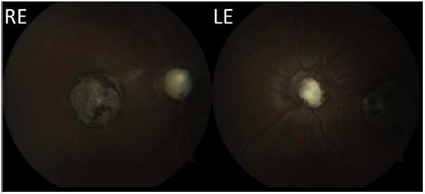
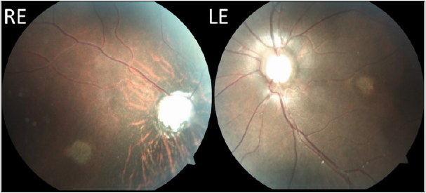
Siblings aged Nine (female) and Five (Male), were referred to the ophthalmology OPD with diminution of vision observed since few months of constant degree. Both children were born to a non-consanguineous marriage and had normal birth and developmental history. Both were known case of Type 1 Diabetics under treatment. On ophthalmologic examination, the elder female child had accommodative esotropia of about 45 degrees. Best Corrected Visual Acuity (BCVA) was counting fingers at 1 meter in both the eyes. The patient also had microphthalmos and microcornea. On retinoscopy, she had a refractive error of -13 dioptre of myopia along with 1.75 dioptre of myopic astigmatism at 45 degrees. The fundus of both eyes showed a pale disc with a large deep cup which was broader inferiorly suggestive of optic disc coloboma. A large single circular scar of about three-disc diameter in size at the macula which was hypopigmented and had small patches of hyperpigmentation in it suggestive of an old posterior uveitis secondary to toxoplasmosis (Figure 1). Also, the patient had coloboma of the choroid inferiorly. Intraocular Pressure was normal in both eyes. The younger male sibling had accommodative esotropia of about 70 degrees. Best Corrected Visual Acuity (BCVA) was only hand movements. The patient also had microphthalmos and microcornea. On retinoscopy, he had a refractive error of -5 dioptres of myopia in the right eye while the left eye was near emmetropia. Patient also had horizontal nystagmus. The Fundus of both eyes showed a pale disc with peripapillary hyperpigmentation, radially arranged vessels, disc excavation with abnormal tissue in its centre-suggestive of morning glory syndrome (Figure 2). Furthermore, the patient had small, half disc diameter hypopigmented patch in both eyes at the macula presumed to be a milder version of the same condition of the elder sister. The patient also had coloboma of the choroid inferiorly. Intraocular Pressure was normal in both eyes. Both patients had no diabetic retinopathy changes and were found to be normal systemically. Both patients had normal mental growth. The parents had normal anterior and posterior segment findings and no similar history was present in the family. Both the children were referred to higher institution for Low Vision Aids (LVA) and on follow-up were found to be doing fine in special school.
Figure 1:Fundus photographs of Right Eye (RE) and Left Eye (LE) of the elder sibling.

Figure 2:Fundus photographs of Right Eye (RE) and Left Eye (LE) of the younger sibling.

Discussion
Congenital optic disc anomalies associated with normal or large optic disc include megalopapilla, morning glory syndrome, optic disc coloboma, optic disc pit and tilted disc. Visual prognosis can range from asymptomatic to total blindness [1]. They are distinguished on fundus examination by an abnormal disc size, abnormalities in disc conformation, and the presence of abnormal tissue within or around the disc and typical arrangement of vessels [2]. Visual prognosis is variable. The morning glory disc abnormality is a congenital condition in which an enlarged, pale, concave disc is surrounded by chorioretinal pigmentary changes and radial spoke vessels said to resemble a morning glory flower [3]. The central portion of the disc is occupied by a white clump of glial tissue. This defect causes a variable degree of visual deficiency, due to associated foveal aplasia [2]. Ocular colobomas are thought to be secondary to defects in embryogenesis in which there is impaired closure of the optic fissures resulting in a defect in the lens, optic nerve, and/or uvea [4]. In consanguineous families, the likelihood of recessive inheritance such as colobomas and Septo-optic dysplasia is significantly higher [5] but in our case report, both siblings were born out of a non-consanguineous marriage. A study [6] conducted at a tertiary eye hospital in western India by Paranjape et al. [6] found that the mean age of children attending Paediatric Ophthalmology OPD was six and half years and concluded that by this time most children would have developed severe disease and lost many years of quality life. Although type 1 diabetes can occur at any age, it peaks in presentation between five–seven years of age and at or near puberty [7]. In our case study the children had been diagnosed with Type 1 diabetes at the age of four years and may have been pointed towards other congenital abnormality.
Conclusion
After extensive research into scientific search engine of the National Center for Biotechnology Information, U.S. National Library of Medicine (PubMed; under English language), we failed to find any previous similar case reports. This seems to be unique case report of Type 1 diabetics sibling having microphthalmos, microcornea, optic disc abnormality associated with macular scars secondary to presumed choroiditis and inferior coloboma of the choroid and high myopia. The case report highlights the lack of awareness in a developing country like India among both doctors and patients regarding necessity of thorough ophthalmic examination at a younger age. Most patients when found to be grossly normal in appearance are not seen by an ophthalmologist till a very late age. Children with type 1 diabetes at a very young age (less than 5 years) need thorough evaluation to rule out any coexisting congenital disorders.
References
- Daroff R (2012) Bradley's neurology in clinical practice. In: Jankovic J, Mazziotta J, Pomeroy S (Eds.), (7thedn), Elsevier, USA, pp. 1-2348.
- Golnik KC (1998) Congenital optic nerve anomalies. Curr Opin Ophthalmol 9(6): 18-26.
- Sobol W, Bratton A, Rivers M, Weingeist T (1990) Morning glory disk syndrome associated with subretinal neovascular membrane formation. Am J Ophthalmol 110(1): 93-94.
- Gregory ECY, Williams MJ, Halford S, Gregory EK (2004) Ocular coloboma: A reassessment in the age of molecular neuroscience. J Med Genet 41(12): 881-891.
- Emma AW, Mehul TD (2010) Septo-optic dysplasia. European Journal of Human Genetics 18(4): 393-397.
- Paranjpe R, Mushtaq I, Thakre A, Sharma A, Dutta D, et al. (2016) Awareness of childhood blindness in parents attending paediatrics ophthalmology outpatient department. Med J DY Patil Univ 9(4): 451-454.
- Harjutsalo V, Sjoberg L, Tuomilehto J (2008) Time trends in the incidence of type 1 diabetes in Finnish children: A cohort study. Lancet 371(9626): 1777-1782.
© 2021 Anuj Kumar Singal. This is an open access article distributed under the terms of the Creative Commons Attribution License , which permits unrestricted use, distribution, and build upon your work non-commercially.
 a Creative Commons Attribution 4.0 International License. Based on a work at www.crimsonpublishers.com.
Best viewed in


.jpg)


 Editorial Board Registrations
 Submit your Article
 Refer a Friend
 Advertise With Us
.jpg)


.jpg)


.bmp)
.jpg)
.png)
.jpg)


.jpg)


.png)

.png)


.png)


