- Submissions

Full Text
Medical & Surgical Ophthalmology Research
Osler’s Sign-The Hidden Clue
Anubhav C* and Kulbhushan PC
Department of Ophthalmology, India
*Corresponding author: Anubhav C and Kulbhushan PC, Department of Ophthalmology, India
Submission: November 27, 2018;Published: December 17, 2018

ISSN 2578-0360 Volume2 Issue4
Abstract
A 55-year-old male presented with a history of joint pains for the past few years. The patient was receiving treatment for arthritis from some other institute. He attended the ophthalmology OPD for a routine ocular checkup, which revealed classical ‘Oslers sign’ specific to Alkaptonuria (AKU). Physical examination also revealed pigmentary changes in the skin. Complete workup lead to the diagnosis of AKU. The joint manifestations were a part of this inborn error of metabolism and not routine arthropathies. We report this case to highlight that inborn errors of metabolism can go undetected for many years if a high degree of suspicion is not there on part of the treating physician.
Keywords:Alkaptonuria; Ocular; Systemic
Case Report
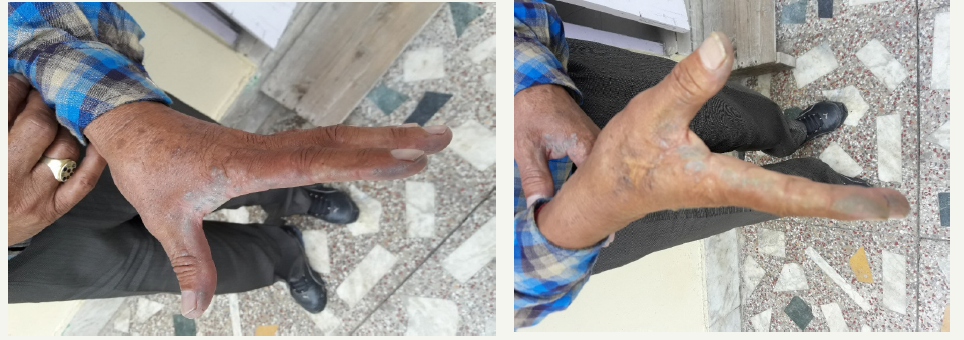
55-year-old male patient presented to the ophthalmology department for routine ocular examination. There was no ocular complaint nor there was a history of any previous ophthalmic consultation. The patient gave a history of having chronic back ache with pains in both his knees and was receiving non-steroidal antiinflammatory drugs from some other institute. There was no other significant medical, surgical, family, traumatic or drug abuse history. Ocular examination was carried out and his visual acuity was 6/6 in both the eyes; pupillary reactions, ocular movements, colour vision, fundus and intraocular pressure were normal bilaterally. Slit lamp and torch examination revealed bluish‐black dots on the cornea, conjuctiva and sclera in the interpalpebral area bilaterally close to the medial and lateral rectus muscles (Figure 1). Dilated conjunctival vessels were seen to be supplying the pigmented areas. There were also pigmentary changes seen on his hands (Figure 2).
Figure 1

Figure 2

On further questioning the patient, a history of darkening of urine on standing was given. A suspicion of AKU arose and we subjected the patient to a battery of investigations. Complete blood count, blood sugar and renal function tests were normal. Urine turned dark on standing in atmospheric air for a few hours. Diagnosis was confirmed by the detection of elevated homogentisic acid level in the urine. Patient was started on Tablet Vitamin C, 500mg twice daily plus he was attached to other clinical departments to rule out any systemic involvements associated with this disease.
Discussion
AKU was the first disease in which a Mendelian recessive inheritance pattern was proposed. The term “alcapton” was first used in 1859 by Boedecke. In the year 1908, Garrod coined the term “inborn error of metabolism” and stated that alkaptonuria occurs due to an enzyme deficiency [1]. It is a rare inherited genetic disorder of tyrosine metabolism characterized by the triad of homogentisic aciduria, ochronosis and arthritis. This condition affects 1 in 250,000 to one million people worldwide. Cases of AKU have been reported from Slovakia and other european countries. It occurs as a result of deficiency of homogentisic acid oxidase (HGAO) which causes the conversion of homogentisic acid (HGA) to maleyl acetoacetate, fumaric acid and acetoacetic acid. In the absence of the HGAO, HGA and benzoquinone acetic acid (BQA) accumulate in the connective tissue(cartilage/collagen) while some part is excreted in the urine. HGA on exposure to air is oxidized to a brown/ black pigment and hence a fresh urine sample of an alkaptonuria patient appears normal but starts darkening on exposure to the air [2].
One of the first symptoms of alkaptonuria is darkening of urine on standing. Osler’s sign i.e scleral pigmentation is usually seen around the 3rd decade of life while the skin pigmentation becomes obvious in the 4th decade and is predominantly seen on the sun exposed areas. One of the first sites to be affected is the ear cartilage. Ochronotic arthropathy starts around the 4th decade and predominantly involves the knees, intervertebral joints in the spine, shoulder joints with narrowing of joint spaces and disc calcifications. Pigment deposits can be seen in the larynx, tonsils, esophagus, dura mater, eardrums, trachea, and bronchi. After the age of 50 years, cardiac findings are in the form of aortic or mitral valvulitis, calcification of coronary arteries and atherosclerotic plaques. Pigment deposits can form stones in the prostate, urethra, and kidneys. The endocrine organs, central nervous system and teeth can also be affected [3].
Life expectancy in AKU patients is normal; however, associated morbidity can be significant. The most common ocular manifestations are bluish‐black discoloration of the conjunctiva, cornea, and sclera. Dilated conjunctival vessels can be present and they supply the pigmented areas on the conjunctiva [4]. The term hereditary ochronosis is applied for the tissue manifestation of the disease. Ocular signs are present in at least 2/3 of the patients affected by hereditary ochronosis. Due to their characteristic appearance, “oil-drops” sign is considered pathognomonic to ochronosis. Other ocular structures involved are the posterior surface of the lens, iris, vitreous, and optic disc. Association with glaucoma, uveitis [5] and epiretinal membrane [4] has also been reported. In the eye, the pigmentation is usually seen at the site of insertion of the medial and lateral rectus muscles. The pigment involves the entire thickness of the sclera and can be either extracellular in location along with collagen fibers, or intracellular within macrophages and fibrocytes [6]. AKU like ochronosis can also be seen due to substances like phenol, hydroquinone, quinine, hydroquinone, minocycline and methyl dopa. The gold standard test for AKU is to detect homogentisic acid in the urine. Various tests are as follows:
a. Alkali test- on addition of NaOH to urine, it turns black.
b. Ferric chloride test- transient green color is seen.
c. Ammonical silver nitrate test-forms black precipitate of silver.
d. Benedict’s test-shows black supernatant and red brown precipitate of cuprous oxide at the bottom.
e. N-butane test-pink brown color is formed.
For bone and joint involvement, X ray of spine and major joints can detect degenerative arthropathies. Further radiographs and ultrasonography can also pick up kidney stones. Magnetic resonance imaging can show thickening of Achilles tendon. Computed Tomography and echo cardiographic studies can reveal cardiovascular abnormalities [7]. Other differential diagnosis includes Nevus of Ota (naevus fuscocaeruleus ophthalmomaxillaris). It is a rare disorder of periocular hyperpigmentation associated with scleral melanosis. The lesions are predominantly unilateral, patchy, bluish gray discoloration of the skin of the face supplied by the first and second divisions of the trigeminal nerve with involvement of the periorbital region, temple, forehead, malar area, and nose. It is predominantly seen in females [8].
Currently as there is no effective therapy for AKU. Treatment modalities includes drugs, physiotherapy, joint replacement surgery, and pain control. Vitamin C is used and is believed to reduce the conversion of HGA to BQA, but some studies consider it to be an unsuitable treatment because of systemic side effects in AKU patients. Some authors advocate low protein diet, liver transplantation, triketone herbicide Nitisinone (that inhibits 4-hydroxyphenyl pyruvate, an enzyme involved in the conversion of hydroxyphenylpyruvate to HGA) and enzyme replacement therapy for the treatment of AKU, but all the therapies have there disadvantages as well [9].
Conclusion
The authors consider that the distinct ocular sites for these lesions could be as a result of vascularity of the area, distinct collagenase, oxygen availibility to polymerize the black tarry pigment, milking action of the upper and lower lids, movement and curvature of the eyeball, limbal anatomy, gravity and the horizontal recti muscle insertion areas where muscular arteries and anterior ciliary vessels form a dense vascular plexus and hence have the dense black pigmentary deposits. Oslers sign is a term also used in pseudo hypertension, graves disease and bacterial endocarditis. AKU is rare disease, therefore the likelihood that this disease going undetected is high. Therefore, a high index of suspicion and awareness on the part of the physician is essential to clinch the diagnosis.
References
- Reddy OJ, Gafoor JA, Suresh B, Prasad PO (2014) Alkaptonuria with review of literature. J NTR Univ Health Sci 3(2): 125-129.
- Nafees M, Muazzam M (2007) Alkaptonuria-case report and review of literature. Pak J Med Sci 23(4): 650-653.
- Tharini GK, Ravindran V, Hema N, Prabhavathy D, Parveen B (2011) Alkaptonuria. Indian J Dermatol 56(2): 194-196.
- Damarla N, Linga P, Goyal M, Tadisina SR, Reddy GS, et al. (2017) Alkaptonuria: A case report. Indian J Ophthalmol 65(6): 518-521.
- Lindner M, Bertelmann T (2014) On the ocular findings in ochronosis: a systematic review of literature. BMC Ophthalmology 14(1): 12.
- Kumar A, Karthikeyan K, Vyas MT (2015) Oslers sign revisited. Indian Dermatol Online J 6(4): 308-309.
- Chauhan S, Garg A, Tegta GR, Thakur K (2017) Skin pigmentation, a window to diagnose Alkaptonuria: a very rare entity. Int J Res Med Sci 5(6): 2801-2805.
- Kumari R, Thappa DM (2006) Familial nevus of ota. Indian J Dermatol 51(3): 198-199.
- Mistry JB, Bukhari M, Taylor AM (2013) Alkaptonuria. Rare Diseases 1: e27475.
© 2018 Anubhav C. This is an open access article distributed under the terms of the Creative Commons Attribution License , which permits unrestricted use, distribution, and build upon your work non-commercially.
 a Creative Commons Attribution 4.0 International License. Based on a work at www.crimsonpublishers.com.
Best viewed in


.jpg)


 Editorial Board Registrations
 Submit your Article
 Refer a Friend
 Advertise With Us
.jpg)


.jpg)


.bmp)
.jpg)
.png)
.jpg)


.jpg)


.png)

.png)


.png)


