- Submissions

Full Text
Modern Research in Dentistry
Mandibular Distractor for Experimental Applications
Khalid Abdullah Alruhaimi1* and Abdullah K Alruhaimi2
1Department of Maxillofacial Surgery, College of Dentistry, King Saud University, Saudi Arabia
2General Practitioner, Riyadh, Saudi Arabia
*Corresponding author: Khalid Abdullah Alruhaimi, BDS, MSc, Dr. Med. Dent, Professor, Department of Maxillofacial Surgery, College of Dentistry, King Saud University, Riyadh, Saudi Arabia
Submission: January 10, 2022;Published: February 08, 2022

ISSN:2637-7764Volume7 Issue2
Abstract
The paper presents an innovation of a mandibular distractor device. It is designed to facilitate applied experimental research in the field of distraction osteogenesis process. The device is configured for attachment to opposing sides of a mandible performing distraction osteogenesis in both sides of the mandible with one distractor.
Keywords: Distraction osteogenesis; Distractor device; Maxillofacial distractor; Distractor for experimental application
Introduction
Distraction osteogenesis is a process of lengthening bone in a gradual manner by distracting or separating one surgically sectioned bony part from an adjacent surgically sectioned bony part with the use of a distractor device. The distraction is typically performed in small daily increments, and generally results in the formation of new bone between the separated bony parts [1,2]. The procedure is used to lengthen short bones or generate new bone in a defected or deficient bony site without the need for a bone graft. Experimental animals such as rabbits are used to study the process of distraction osteogenesis and investigate methods to enhance the speed or quality of the gained bone by adding various materials [3-6].
Material and Methods
Description of the distractor
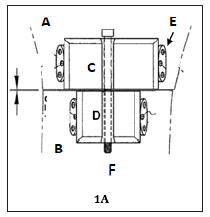
The distractor device includes a distractor body and an activation bar that extends through a central portion of the distractor body. The distractor body is defined by an anterior plate and a posterior plate, each of which are attachable to opposing sides of the mandible with self-drilling mini screws. The anterior plate includes a tubular portion with a threaded interior wall. The posterior plate includes a tubular portion with a smooth interior wall. The activation bar includes a threaded portion and a smooth portion. The activation bar can be disposed within the body such that the threaded portion of the bar is threaded to the interior wall of the anterior plate, while the smooth portion of the bar extends within the smooth interior wall of the posterior plate. The smooth portion of the bar and the interior wall of the posterior plate permit free rotation of the posterior portion of the bar within the posterior plate during rotation of the anterior threaded part of the bar into the threaded anterior part of the device. The start of the activation process of the device becomes feasible by a clockwise rotation of the activation bar so it is disengaged from the anterior plate. The anterior plate and the anterior mandibular part attached thereto move anteriorly and further away from the posterior plate and the posterior part of the mandible attached thereto which remains in position (Figure 1A).
Surgical application
A medical practitioner can form a cortectomy line on the lower border and on the lateral and medial surfaces of the rabbit’s mandible as illustrated in Figures 1A. The cortectomy line divides the rabbit’s mandible into two parts. The body is then positioned on the lower border of the rabbit’s mandible so that the anterior plate of the device is secured to one side of the cortectomy line and the posterior plate is secured on the other side of the cortectomy line. Each attachment tab is then attached within the lateral surface of each side of the rabbit’s mandible with appropriate fasteners. The anterior plate of the body can be secured to opposing sides of the anterior mandibular part of the rabbit’s mandible and the posterior plate of the body can be secured to opposing sides of a posterior mandibular part of the rabbit’s mandible.
The practitioner can then separate the two sections of the rabbit’s mandible by wedging a chisel along the cortectomy line (Figure 1A). The practitioner can then utilize a screwdriver to activate the device, i.e., rotate the activation bar in a clockwise direction, and thereby move the anterior plate anteriorly along with the sectioned anterior part of the mandible, as illustrated in Figure 1B. Every full rotation of the activation bar can move the anterior plate (and the mandibular part attached thereto) 0.5mm of distraction distance. It should be understood that activation of the device moves the anterior plate and not the posterior plate. In other words, the posterior plate is not moved for the distraction. The distraction increments can be 1mm per day, in accordance with international protocol for daily distraction.
Figure 1A: A top view of a rabbit’s mandible and the mandibular distraction device attached thereto. Showing view, the osteotomy line prior to formation of the mandibular gap (arrowed) between the posterior(a) and anterior (b) sectioned parts of the mandible. Note the posterior part of the distractor body (c) is wider than the anterior part (d) to accommodate the divergence of the two sides of the rabbit’s mandible. (E) four anchor plates fastened to the lateral sides of the opposing mandibles with mini-screws. Two plates are attached to each side of the posterior distractor part and another two plates attached to each side the anterior distractor part. (F) is the tip of the activation bar.

Figure 1B: Showing view of activation of the distractor device led to formation of mandibular distraction space (arrowed).

Discussion
The common available distractors used on the experimental models act on the mandible unilaterally [5-7]. The unilateral device includes separate distractors, one for each side of the mandible. Subsequently, each unilateral distraction device has to be activated separately each day. Further, two separate distractors involve longer surgical time, longer daily activation time and more surgical armamentarium costs. Thus, one mandibular distractor device is solving the a fore mentioned problems [1,2]. The present device on the other hand can be secured to both sides of the mandible. As such, the device can be activated for distracting both sides of the mandible at one time. To activate the device, the activation bar is rotated to unthread or disengage the activation bar from the interior wall of the anterior plate. For example, the device is rotated in daily increments to incrementally disengage the threaded portion of the activation bar from the threaded interior wall of the anterior plate. Rotation of the activation bar in this manner incrementally moves the anterior plate and the mandibular part attached thereto anteriorly with one device, while the posterior plate and the posterior part of the mandible remains in position [8,9].
Conclusion
The common available distractors act on the mandible unilaterally. The unilateral device includes two separate distractors, one for each side of the mandible. Subsequently, each unilateral distraction device has to be activated separately each day. Additionally, the activation bar is exposed at each side of the mandible. The presented mandibular distractor device is configured for attachment to both of the opposing sides of a mandible, e.g., a rabbit’s mandible, such as for experimental studies for performing distraction osteogenesis. The device can be secured to both sides of the mandible. As such, the device can be activated for distracting both sides of the mandible at one time.
Acknowledgment
Author would like to thank the College of Dentistry Research Center and Deanship of Scientific Research at King Saud University, Saudi Arabia for funding this research project (research project # 0601).
References
- Alruhaimi K (2017) Mandibular distractor device, pp. 1-6.
- Alruhaimi K (2000) A submerged osteodistraction device: an innovative technique for experimental animal studies. J Craniofac Surg 11(1): 59-61.
- Alruhaimi K (2001) Comparison of different distraction rates in the mandible: an experimental investigation. Int J Oral Maxillofac Surg 30(3): 225-228.
- Alruhaimi K (2001) Effect of calcium sulfate on the rate of osteogenesis in distracted bone. Int J Oral Maxillofac Surg 30(3): 228-233.
- AlSebaei MO, Gagari E, Papageorge M (2005) Mandibular distraction osteogenesis: A rabbit model using a novel experimental design. J Oral Maxillofac Surg 63(5): 664-672.
- Aida T, Yoshioka I, Tominaga K, Fukuda J (2003) Effects of latency period in a rabbit mandibular distraction osteogenesis. Int J Oral Maxillofac Surg 32(1): 54-62.
- Meng Q, Chen G, Long X, Deng M, Cai I, et al. (2011) Histological evaluation of condylar hyperplasia model of rabbit following distraction osteogenesis of the condylar neck. J Oral Rehabilit 38(1): 27-33.
- Mutlu I, Aydintug YS, Kaya A, Bayar GR, Suer BT, et al. (2012) The evaluation of the effects of hyperbaric oxygen therapy on new bone formation obtained by distraction osteogenesis in terms of consolidation periods. Clin Oral Investig 16(5): 1363-1370.
- Wei H, Zili L, Yuanlu C, Biao Y, Cheng L, et al. (2011) Effects of icariin on bone formation during distraction osteogenesis in the rabbit mandible. Int J Oral Maxillof Surg 40: 413-418.
© 2022 Khalid Abdullah Alruhaimi. This is an open access article distributed under the terms of the Creative Commons Attribution License , which permits unrestricted use, distribution, and build upon your work non-commercially.
 a Creative Commons Attribution 4.0 International License. Based on a work at www.crimsonpublishers.com.
Best viewed in


.jpg)


 Editorial Board Registrations
 Submit your Article
 Refer a Friend
 Advertise With Us
.jpg)


.jpg)


.bmp)
.jpg)
.png)
.jpg)


.jpg)


.png)

.png)


.png)


