- Submissions

Full Text
Modern Research in Dentistry
Decreasing the Risk of Incisor Root Resorption during the Orthodontic Management of Palatally Impacted Canines: Case Report
Emad Hussein*, Sari Amer, Stathis Efstathiou, Mohammad A Mowais, Nezar Watted, Khaled Qattawi and Manal Samarah
Clinical master program of orthodontics, Arab American University of Palestine, Palestine
*Corresponding author: Emad Hussein, Professor, Clinical master program of orthodontics, Arab American University of Palestine, Palestine
Submission: November 05, 2021;Published: November 29, 2021

ISSN:2637-7764Volume7 Issue1
Abstract
Palatally impacted canines can lead to root resorption of adjacent incisors; root resorption can also result due to orthodontic treatment when unplanned orthodontic movements occur close to the incisors roots. Care should be taken during the first stages of tooth movements in order to avoid root resorption. The first tooth movement of the impacted canine should aim to take the canines away from the roots of incisors. This case report represents an example how we avoided root resorption of teeth adjacent to the impacted canine by taking the canines away by a modified transpalatal bar.
Keywords: Canine impaction; Root resorption; Modified bar
Introduction
The second most frequently impacted teeth are the canines, just behind the third molars
[1]. Many studies showed that the incidence of maxillary canine impaction ranges between
0.24% to 3%, which varies depending on the ethnicity [2-5], this condition affects females
2 to 3 times more frequently than males [2,6]. According to the literature, 85% of canine
impactions occur palatally and 15% buccally [7]. Many studies found that unilateral impaction
ranges from 60-92% [8-11].
Palatally impacted canines can lead to root resorption of adjacent incisors; root resorption
can also result due to orthodontic treatment when unplanned orthodontic movements occur
close to the incisors roots [12-16]. Care should be taken during the first stages of tooth
movements in order to avoid root resorption. The first tooth movement of the impacted
canine should aim to take the canines away from the roots of incisors [17-20]. The movement
should be directed distally and lead to vertically up righting the canine in the palate. In this
article, we will discuss a case of bilaterally impacted canines treated by surgical exposure with
orthodontic traction and alignment by the use of a modified double Transpalatal bar [21-25].
Case Report
S.K is a 16-year old female who presented with the main complaint of impacted maxillary canines as informed by her dentist. She presented with a symmetric face and competent lips (Figure 1A). Upon smiling, she showed a reduced appearance of anterior teeth under lip line, and a 3mm midline diastema (Figure 1B). S.K had a straight profile with increased lower anterior height. This patient had a Class III skeletal discrepancy due to a hypoplastic maxilla and a slightly prognathic mandible. The flared upper incisors and the perioral soft tissue masked the skeletal discrepancy (Figures 1C & 1D) and (Figure 1K). Intraorally, she had proclined upper anterior teeth with a midline diastema, increased overjet and average overbite. The deciduous maxillary canines were retained. Palatal bulges were present on both sides of palatal midline indicating palatally impacted canines (Figures 1E-I).
S.K had a Class II molar relationship with slight maxillary constriction; a crossbite was present between the upper right deciduous canine and lower right first premolar. 1mm of space was evident in the lower arch (Figure 1E-I). The panoramic x ray shows the presence of all teeth including wisdom teeth; the maxillary canines were impacted behind the lateral incisors and crossing the lateral incisors pulp canal line (Figure 1J).
Figure 1( A-K): Pretreatment orthodontic records, extra and intra-oral pictures, panoramic and lateral skull x-rays.

Treatment plan
Treatment plan of this case aimed to create enough space for the maxillary impacted canines and to bring the impacted canines to the arch, to correct the anterior teeth inclination, and to accept the dentoalveolar compensation of the skeletal discrepancy.
Treatment progress
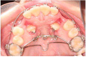
A palatal appliance “double transpalatal bar” was designed, which is comprised of a Transpalatal bar and a 3-helix bar. Both bars were soldered on first molar bands Figure 2. The purpose of the 3-helix bar is an anchorage for the elastic thread that pulls the impacted canines away from the roots of the incisors. After vertical eruption of canines, the 3-helix bar is cut, and the second transpalatal bar is left in place for anchoring the maxillary first molars (Figure 3A).
Figure 2: Modified transpalatal bar with the impacted canines being uprighted through the attachments from the lingual surface to the bar-helices.
Pinnacle®.022” orthodontic bracket appliances from Ortho
Technology® South Carolina, USA with their version of the
McLaughlin, Bennett, and Trevisi prescription were bonded;
alignment and leveling were performed; deciduous canines were
extracted, and space was opened to accommodate the erupting
canines (Figures 3A-E).
Surgical exposure was carried out using a closed technique.
Eyelets (Ortho Technology) were bonded during surgery and
Elastic Threads (Ortho Technology), pulling the impacted canines
from the roots of the incisors started, were fixed on the helices. Flap
was closed and changing the elastic thread was carried out every
three weeks (Figures 3A-E).
Figure 3 (A-E): Labial attachment of the impacted canines after up righting them away from incisors’ roots.

After canines were vertically up-righted and away from the incisors, bonding of two eyelets on the labial sides of these canines was performed, and traction of the canines to the arch started (Figure 3D) and (Figures 4A-E). When canines were close to the arch, .016” TruFlex™ Nickel Titanium Archwire (Ortho Technology) wire was used to align the canines and to bring them out of crossbite (Figure 5A-C), (Figures 6A-C) and (Figures 7A-C). During a later stage, eyelets were removed and replaced by canine brackets (Figure 8A-H).
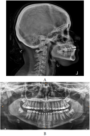
Leveling and alignment continued and torque of the upper incisors was adjusted. Finally, maxillary canines were placed in position with proper angulation and torque (Figures 9A-C) and (Figure 10A&B).
Figure 4 (A-E): Continued traction of the impacted canines to the arch by elastic thread from the main archwire in a direction to their prepared places.

Figure 5 (A-C): Alignment of the canines by direct engagement of the NiTi wire through the eyelets.
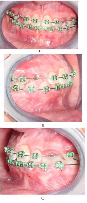
Figure 6 (A-C): Continued canines alignment by the NiTi wire.
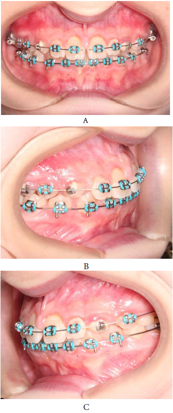
Figure 7 (A-c): The canines are aligned by the eyelet- NiTi wire before replacing the eyelets by brackets.
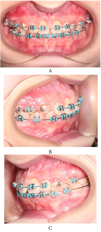
Figure 8 (A-H): Leveling and alignment completed spaces mesial and distal to the upper lateral incisors maintained to correct the Bolton discrepancy.

Figure 9(A-C): Settling the occlusion by vertical elastics.
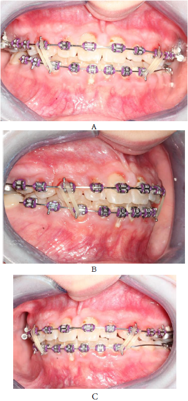
Figure 10 (A,B): Panoramic and lateral skull x-rays before debonding
Discussion and Conclusion
Various complications can occur in the presence of palatally
impacted canine [26,27]. Root resorption of the adjacent incisors is
common sequelae that should be well considered in the treatment
plan. Earlier studies by Ericson and Kurol using conventional
radiography, estimated that in 12.5% of the ectopically maxillary
canines caused some resorption of the adjacent incisors [12].
Later on, with the emergence of CT as an essential diagnostic
tool, Ericson reported that root resorption had occurred in 38% of
the lateral incisors and 9% of the central incisors [28], while other
studies reported 25% - 67.7% root resorption for lateral incisor and
5%-18% for the central incisor [28-30]. 50% more root resorption
detection was reported when using CT compared to intraoral x
rays [28]. A recent study, using CBCT to determine the main factors
associated with the palatal displaced canines and incisor root
resorption, concluded that canines in contact with adjacent incisor
roots was the only risk factor for incisor root resorption, while
canine angulation and proximity to midline were not considered
factors affecting root resorption [31].
The treatment of impacted canines can be by either the surgical
exposure followed by orthodontic traction of the palatally impacted
canine, or the extractions of the impacted canines and leaving the
first premolar in place of the impacted canine.
The most important step during orthodontic treatment of a
palatally impacted canine is to start by taking the impacted canine
away from the roots of the incisors after correct diagnosis and
surgical exposure; this can be achieved by up righting the palatally
impacted canine vertically in the palate.
Several methods were described for traction of the canines,
using a Ballista spring and pulling them from a Transpalatal bar and
more recently the use of miniscrews to achieve this purpose.
We suggest the use a combined Transpalatal bar and a 3-helical
bar appliance, the helices will be the site of attachment of the elastic
thread to pull the canines away from the incisors and when the
canines are up righted, the helical bar is removed, and a TPB is left
in place for anchorage purposes.
References
- Kalle A, Lehtinen R, Oksala E (1972) An orthopantomography study of prevalence of impacted teeth. International Journal of Oral Surgery 1(3): 117-120.
- Dachi SF, Howell FV (1961) A survey of 3,874 routine full-mouth radiographs: I. A study of retained roots and teeth. Oral Surgery Oral Medicine Oral Pathology 14(8): 916-924.
- Thilander B, Myrberg N (1973) The prevalence of malocclusion in Swedish schoolchildren. Scand J Dent Res 81(1): 12-21.
- Takahama Y, Aiyama Y (1982) Maxillary canine impaction as a possible microform of cleft lip and palate. The European Journal of Orthodontics 4(4): 275-277.
- Raffaele S, Baccetti T (2004) Dentoskeletal features associated with unilateral or bilateral palatal displacement of maxillary canines. The Angle Orthodontist 74(6): 725-732.
- Wilbur JD (1969) Treatment of palatally impacted canine teeth. Am J Orthod 56(6): 589-596.
- Hitchin AD (1951) The impacted maxillary canine. Dent Pract Dent Rec 2(4):100-103.
- Power SM, Short Mary BE (1993) An investigation into the response of palatally displaced canines to the removal of deciduous canines and an assessment of factors contributing to favourable eruption. British Journal of Orthodontics 20(3): 215-223.
- Sune E, Kurol J (1987) Radiographic examination of ectopically erupting maxillary canines. Am J Orthod Dentofacial Orthop 91(6): 483-492.
- Bishara SE (1992) Impacted maxillary canines: a review. American Journal of Orthodontics and Dentofacial Orthopedics 101(2): 159-171.
- Abron A, Mendro RL, Kaplan S (2004) Impacted permanent maxillary canines: diagnosis and treatment. NY State Dent J 70(9): 24-28.
- Ericson S, Kurol PJ (2000) Resorption of incisors after ectopic eruption of maxillary canines: a CT study. The Angle Orthodontist 70(6): 415-423.
- Lai CS, Bornstein MM, Mock L, Heuberger BM, Dietrich T, et al. (2013) Impacted maxillary canines and root resorptions of neighbouring teeth: a radiographic analysis using cone-beam computed tomography. European Journal of Orthodontics 35(4): 529-538.
- Olsen SH, Pirttiniemi P, Kerosuo H, Limchaichana NB, Pesonen P, et al. (2015) Root resorptions related to ectopic and normal eruption of maxillary canine teeth-A 3D study. Acta Odontologica Scandinavica 73(8): 609-615.
- Alqerban A, Jacobs R, Fieuws S, Willems G (2016) Predictors of root resorption associated with maxillary canine impaction in panoramic images. Eur J Orthod 38(3): 292-299.
- Alemam AS, Alhaija ESA, Mortaja K, AlTawachi A (2020) Incisor root resorption associated with palatally displaced maxillary canines: Analysis and prediction using discriminant function analysis. American Journal of Orthodontics and Dentofacial Orthopedics 157(1): 80-90.
- Harry J (1983) The etiology of maxillary canine impactions. American Journal of Orthodontics 84(2): 125-132.
- Becker A, Smith P, Behar R (1981) The incidence of anomalous maxillary lateral incisors in relation to palatally-displaced cuspids. The Angle Orthodontist 51(1): 24-29.
- Peck S, Peck L, Kataja M (1994) The palatally displaced canine as a dental anomaly of genetic origin. The Angle Orthodontist 64(4): 249-256.
- Counihan K, Al Awadhi EA, Butler J (2013) Guidelines for the assessment of the impacted maxillary canine. Dental Update 40(9): 770-777.
- Wolf JE, Mattila K (1979) Localization of impacted maxillary canines by panoramic tomography. Dentomaxillofacial Radiology 8(2): 85-91.
- Fox NA, Fletcher GA, Horner K (1995) Localising maxillary canines using dental panoramic tomography. British Dental Journal 179 (11-12): 416-420.
- Chaushu S, Chaushu G, Becker A (1999) The use of panoramic radiographs to localize displaced maxillary canines. Oral Surg Oral Med Oral Pathol Oral Radiol Endod 88(4): 511-516.
- Armstrong C, Johnston C, Burden D, Stevenson M (2003) Localizing ectopic maxillary canines-horizontal or vertical parallax? Eur J Orthod 25(6): 585-589.
- Alqerban A, Hedesiu M, Baciut M, Nackaerts O, Jacobs R, et al. (2013) Pre-surgical treatment planning of maxillary canine impactions using panoramic vs cone beam CT imaging." Dentomaxillofacial Radiology 42(9): 20130157.
- Eslami E, Barkhordar H, Abramovitch K, Kim J, Masoud MI (2017) Cone-beam computed tomography vs conventional radiography in visualization of maxillary impacted-canine localization: A systematic review of comparative studies. American Journal of Orthodontics and Dentofacial Orthopedics 151(2): 248-258.
- Power SM, Short MB (1993) An investigation into the response of palatally displaced canines to the removal of deciduous canines and an assessment of factors contributing to favourable eruption. British Journal of Orthodontics 20(3): 215-223.
- Ericson S, Kurol J (1988) Early treatment of palatally erupting maxillary canines by extraction of the primary canines. European Journal of Orthodontics 10(4): 283-295.
- Parkin N, Furness S, Shah A, Thind B, Marshman Z, et al. (2012) Extraction of primary (baby) teeth for unerupted palatally displaced permanent canine teeth in children. Cochrane Database of Systematic Reviews 12: CD004621.
- Baccetti T, Mucedero M, Leonardi M, Cozza P (2009) Interceptive treatment of palatal impaction of maxillary canines with rapid maxillary expansion: a randomized clinical trial. American Journal of Orthodontics and Dentofacial Orthopedics 136(5): 657-661.
- Leonardi M, Armi P, Lorenzo F, Baccetti T (2004) Two interceptive approaches to palatally displaced canines: a prospective longitudinal study. The Angle Orthodontist 74(5): 581-586.
© 2021 Emad Hussein. This is an open access article distributed under the terms of the Creative Commons Attribution License , which permits unrestricted use, distribution, and build upon your work non-commercially.
 a Creative Commons Attribution 4.0 International License. Based on a work at www.crimsonpublishers.com.
Best viewed in


.jpg)


 Editorial Board Registrations
 Submit your Article
 Refer a Friend
 Advertise With Us
.jpg)


.jpg)


.bmp)
.jpg)
.png)
.jpg)


.jpg)


.png)

.png)


.png)


