- Submissions

Full Text
Modern Research in Dentistry
Paraneoplastic Pemphigus: Clinical Case Report and Oral Care Management
Aline Marques Ferreira1*, Letícia Rodrigues Pereira1, Ademilton Couto Nascimento Jr1 and Ana Luiza Ribeiro Oliveira Avi2
1Dental surgeon; Residency in Hospital Dentistry at the Hospital de Amor de Barretos, Barretos, Brazil
2Dental surgeon; Department Dentistry, Hospital de Amor de Barretos, Barretos, Brazil
*Corresponding author: Aline Marques Ferreira, Dental surgeon; Residency in Hospital Dentistry at the Hospital de Amor de Barretos, Barretos, Brazil
Submission: May 26, 2021;Published: June 18, 2021

ISSN:2637-7764Volume6 Issue3
Abstract
Introduction: Paraneoplastic pemphigus is a rare, autoimmune, vesiculobullous disease that affects patients with neoplasia, usually lymphoma or chronic lymphocytic leukemia. It is a very serious condition and related to high rates of morbidity and mortality.
Case report: A patient with a previous diagnosis of chronic lymphocytic leukemia, in disease progression, reported the appearance of ulcers in the oral cavity, multiple bleeding skin lesions, severe pain and persistent fever for 15 days, which started after the use of antibiotics. The clinical examination showed involvement of the ocular, oral and urogenital mucosae, body, scalp, back and palms, with crusty, bleeding and coalescent lesions. The dentistry team worked throughout the hospitalization period and oral care. Conclusion: The diagnosis and care of paraneoplastic pemphigus patients is multidisciplinary. The dentist has a fundamental role in the team, aiming at the patient integral care, providing comfort and a better quality of life.
Keywords: Paraneoplastic pemphigus; Oral lesions; Oral care
Introduction
Paraneoplastic pemphigus is a rare autoimmune, vesiculobullous disease that affects patients with neoplasia, usually lymphoma or chronic lymphocytic leukemia [1]. It is a very serious condition and is related to high rates of morbidity and mortality. Clinically, it may be similar to pemphigus, pemphigoid, erythema multiforme, Stevens-Johnson syndrome, graftversus- host disease, or lichen planus [2]. The lesions appear as erosions, spots, papules or blisters, starting by the mucous membranes, often oral mucositis is the first symptom, and after the appearance of skin lesions, mainly in the upper body [3]. The aim of this study is to describe a case of paraneoplastic pemphigus lesions and oral care provided by the dental surgeon.
Case Report
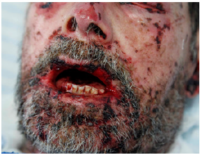
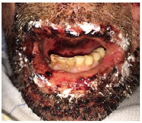
57-year-old male patient, leukoderma, with previous diagnosis of chronic lymphocytic leukemia, in disease progression. He reported the appearance of ulcers in the oral cavity, multiple bleeding skin lesions, severe pain and persistent fever for 15 days, which started after the use of antibiotics. The clinical examination showed involvement of the ocular, oral and urogenital mucosae, body, scalp, back and palms, with crusty, bleeding and coalescent lesions (Figure 1) The first diagnostic hypothesis was Stevens-Johnson syndrome. The patient was initially admitted to the Intensive Care Unit and remained in isolation, as there was a high risk of sepsis. During the hospitalization period, the patient received antibiotic therapy, medications for pain control and clinical care of the wounds. The dentistry team worked throughout the hospitalization period, guiding and performing oral hygiene, caring for oral and peri-oral lesions, with 0.12% Chlorhexidine and moisturizing the lips and wounds with Bepantol® (Figure 2 & 3). Sometimes, photo biomodulation (100mW, 780nm, 3J / cm²), in non-bleeding areas, for analgesia and tissue repair was used. After a punching procedure in an abdomen lesion performed by the medical team, the diagnosis of paraneoplastic pemphigus was confirmed.
Figure 1:bleeding mucocutaneous lesions.

Figure 2:oral cavity after oral hygiene and photo biomodulation therapy.

Figure 3:lip hydration with Bepantol®.

Discussion
Paraneoplastic pemphigus was first identified in 1990 by Anhalt [4]. It is related to malignant or benign neoplasia, which may still be hidden or already diagnosed [1]. In the reported case, the patient had a previous diagnosis of leukemia prior to the manifestation of lesions of the paraneoplastic pemphigus. Hematological neoplasms, including leukemia, are the most related tumors, representing 84%, according to Sehgal and Srivastava [5]. Despite occurring in children, adults from 45 to 70 years are the most commonly affected [1,3]. In this case, the patient was in the most affected age group. Paraneoplastic pemphigus often manifests itself first in the mucous membranes, with erosions, blisters and painful ulcers. In the oral cavity, severe stomatitis is commonly diagnosed. Skin involvement is typically diffuse, and the lesions vary clinically from spots to ulcerations [6,7]. A clinical study of 88 patients, conducted by Ohzono and colleagues, revealed that 93% had oral involvement and 67% had mucocutaneous lesions [8]. In the case described, the patient reported onset of lesions in the oral cavity and that in a few days spread to the skin of the body and arms. The ulcerations presented on mucous membranes were bleeding and extremely painful, which made feeding and oral hygiene difficult. The dentistry team, through the oral care, with photo biomodulation therapy and mucosa hydration, provided symptom relief and oral hygiene with 0.12% chlorhexidine helped in the prevention of secondary infection of the lesions. Immunosuppressant drugs, are often administered to improve mucocutaneous involvement, increase the risk of infectious complications and can lead to sepsis [7].
Thus, as cavity lesions are possible entry for microorganisms, a careful oral hygiene supervised by a dentist is extremely important. This disease treatment remains undefined, but always with the aim of reducing inflammation, suppressing the immune response and providing proper care for the wounds [6]. Paraneoplastic pemphigus is a serious disease, with high rates of morbidity and mortality, therefore, patients require intensive care [3,7].
Conclusion
The diagnosis and management of paraneoplastic pemphigus is multidisciplinary. The dentist has a fundamental role in the team, aiming at the integral care of the patient, providing comfort and a better quality of life.
References
- Tirado Sanchez A, Bonifaz A (2017) Paraneoplastic pemphigus. A life-threatening autoimmune blistering disease. Actas Dermosifiliogr 108(10): 902-110.
- Rashid H, Lamberts A, Diercks GFH, Pas HH, Meijer JM, et al. (2019) Oral lesions in autoimmune bullous diseases: An overview of clinical characteristics and diagnostic algorithm. Am J Clin Dermatol 20(6): 847-861.
- Kappius RH, Ufkes NA, Thiers BH (2021) Paraneoplastic pemphigus. StatPearls. Treasure Island, Florida, USA.
- Anhalt GJ, Kim SC, Stanley JR, Korman NJ, Jabs DA, et al. (1990) Paraneoplastic pemphigus. An autoimmune mucocutaneous disease associated with neoplasia. N Engl J Med 323(25): 1729-1735.
- Sehgal VN, Srivastava G (2009) Paraneoplastic pemphigus/paraneoplastic autoimmune multiorgan syndrome. Int J Dermatol 48(2): 162-169.
- Billet SE, Grando SA, Pittelkow MR (2006) Paraneoplastic autoimmune multiorgan syndrome: review of the literature and support for a cytotoxic role in pathogenesis. Autoimmunity 39(7): 617-630.
- Maruta CW, Miyamoto D, Aoki V, Carvalho RGR, Cunha BM, et al. (2019) Paraneoplastic pemphigus: a clinical, laboratorial, and therapeutic overview. An Bras Dermatol 94(4): 388-398.
- Ohzono A, Sogame R, Li X, Teye K, Tsuchisaka A, et al. (2015) Clinical and immunological findings in 104 cases of paraneoplastic pemphigus. Br J Dermatol 173(6): 1447-1452.
© 2021 Aline Marques Ferreira. This is an open access article distributed under the terms of the Creative Commons Attribution License , which permits unrestricted use, distribution, and build upon your work non-commercially.
 a Creative Commons Attribution 4.0 International License. Based on a work at www.crimsonpublishers.com.
Best viewed in


.jpg)


 Editorial Board Registrations
 Submit your Article
 Refer a Friend
 Advertise With Us
.jpg)


.jpg)


.bmp)
.jpg)
.png)
.jpg)


.jpg)


.png)

.png)


.png)


