- Submissions

Full Text
Modern Research in Dentistry
Cornelia De Lange Syndrome and Dental Treatment: A Case Report
Chaza Kouchaji* and Maram Mohamad Samir al Saitary
Pediatric dental department, Faculty of dental medicine, Damascus university, Syria
*Corresponding author:Chaza Kouchaji, Pediatric dental department, Faculty of dental medicine, Damascus university, Syria
Submission: August 08, 2020;Published: March 16, 2021

ISSN:2637-7764Volume6 Issue2
Abstract
Cornelia De Lange Syndrome is a multisystemic disease with expressing variable physical, cognitive, and behavioral characteristics. It is a genetic disorder that affects many organs, leading to various clinical presentations. In this case presentation we will report a case of CdLS phenotype, along with extra and intra oral finding and radiographic, then dental management and its follow up.
Introduction
History
Previously, in 1849, the anatomist Willem Vrolik (1801-1863) reported a case as an
extreme example of oligodactyly, and the German doctor Brachmann published, in 1916, a
case of symmetric monodactyly, antecubital webbing, dwarfism, cervical ribs, and hirsutism.
Throughout history, other names for the syndrome have included Amsterdam dwarfism,
Bushy syndrome, or Brachmann syndrome [1] CdLS is a multisystemic disease with expressing
variable physical, cognitive, and behavioral characteristics. It is a genetic disorder that affects
many organs, leading to various clinical presentations (1).
It is thought that this syndrome was first described in 1916 by Dr. W. Brachmann, a
Dutch pediatrician after which this syndrome is occasionally named (Brachmann-De Lange
Syndrome). However, the syndrome is most commonly named after Prof. Cornelia De Lange
(1871-1950) who reported her first two cases in 1933 (Figure 1) and presented an account of
this syndrome in 1941 in the meeting of the Neurology Society of Amsterdam (2).
Frequency
This syndrome affects 1 in 10000 to 30000 newborn [2].
Genotypes
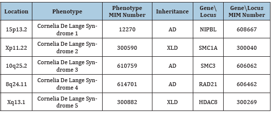
Cornelia de Lange Syndrome (CdLS) has five distinctive genotypes numbered from 1 to 5 with CdLS 1 is the most common which contributes 50-60% of cases. The Table 1 shows the five different genotypes with their mutation locations, phenotype and their MIM numbers in addition to their related genes and genes MIM numbers and their inheritance types [2]. The affected genes (NIPBL, SMC1A, SMC3, RAD21, and HDAC8) all code proteins which are part of the cohesion pathways.
Table 1:Phenotypes of CdLS with their related gene locations and MIM numbers. (www.omim.org).
Clinical manifestations
This syndrome consists of multiple skeletal deformities with
moderate to severe mental retardation associated with facial
dysmorphism. There is usually intrauterine growth retardation
demonstrated by low birth weight [3] as well as postnatal growth
retardation demonstrated by short stature [4]. This can be at least
partially attributed to feeding difficulties in the early childhood.
The latter is one of the most common early demonstrations of the
syndrome and mainly due to Gastro-Esophageal Reflux Disease
(GERD) [4].
The main skeletal abnormalities include: micromilia,
clinodactyly, single transverse palmar crease, proximal implantation
of thumbs, syndactyly of 2nd and 3rd toes.
Regarding the mental retardation, most cases can categorized
as profoundly disabled [5]. the Intellectual Quotient ranges from
below 30 to 86 (average: 53) with initial hypertonicity as the most
stable finding that deter their performance.
Facial dysmorphism findings include microcephaly, highly
arched bushy eyebrows with synophrys [4], long thick eyelashes,
depressed nasal bridge, anteverted nostrils [4], and low set
posteriorly rotated deformed ears [3] with hearing loss [4].
Dental manifestations
Extra-oral manifestations include: long shallow philtrum
with prominent symphysis, down slanted mouth angles, and
microganthia [4].
Intra-oral examination shows usually highly arched palate with
delayed eruption [4]. From the pedodontics perspective, it is worth
to mention main behavioral symptoms. CdLS children are usually
mentally retarded with hypertonicity and hyperactivity with
attention problems. Although they are generally shy but the may
present combative or self-injurious behavior [4,5].
Case Report
In this paper, we report a case of CdLS phenotype, along with extra and intra oral finding and radiographic, then dental management and its follow up.
Presentation
9 years old girl with phonation difficulties and hearing, was
brought to the Pediatric Dentistry Clinic, Damascus University,
Damascus, Syria. The later was treated with unclear surgical
procedure at age of 2 years.as well as a dysmorphic face since birth
which was not fully investigated.
This girl was complaining of continuous pain of 2nd primary
lower molars.
Extra-oral findings
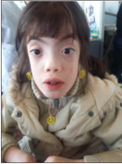
Well noticed face dysmorphism with low frontal hairline, highly arched thick eyebrows, widely spaced eyes (hypertelorism) with mild epicanthal fold, triangular face, anteroverted nostrils, shallow protruded philtrum and wide mouth with downwards slanting corners (Figure 1). No skeletal deformities. We found the facial findings to be suggestive of Cornelia de Lange Syndrome (CdLS).
Figure 1: Wide mouth with downwards slanting corners.
Intra-oral findings
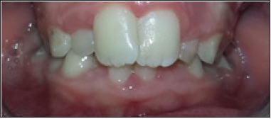
Highly arched palate, significant overlap, narrowed mandibular and maxillary arches, crowded teeth. (Figure 2-4). Type I caries lesions in 54, 64, 74, and 84.
Figure 2:Highly arched palate and significant overlap.
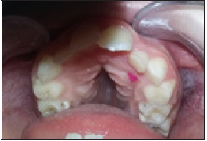
Figure 3: Narrowed mandibular and maxillary arches.
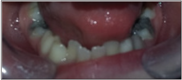
Figure 4: Crowded teeth.
Radiological findings
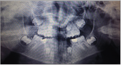
Panoramic X ray showed loss of 2nd premolar buds (Figure 5).
Figure 5: Panoramic X ray showed loss of 2nd premolar buds.
Management
A. Extraction of 74 and 85.
B. Restorative treatment with Glass-Ionomer
Composite fillings of 54, 55, and 64.
C. Root canal treatment with stainless steel crown
under intravenous sedation.
Follow-up
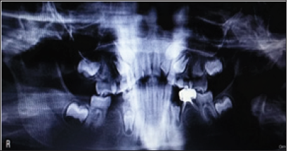
The patient was followed up one month later clinically as the restorations and root canal treatment found satisfactory esthetically and functionally. The extractions site showed good healing and uncomplicated. Also, a follow up panoramic X ray was performed (Figure 6). Which showed no X ray transparencies?
Figure 6:A follow up of panoramic X ray.
References
- Cascella M, Muzio MR (2020) Cornelia de Lange Syndrome. Book from StatPearls Publishing, Treasure Island, Florida, USA.
- Boyle MI, Jespersgaard C, Brøndum Nielsen K, Bisgaard AM, Tümer Z (2015) Cornelia de Lange Syndrome. Clin Genet 88(1): 1-12.
- Hei M, Gao X, Wu L (2018) Clinical and genetic study of 20 patients from China with Cornelia de Lange Syndrome. BMC Pediatr 18(1): 64.
- Kline AD, Grados M, Sponseller P, Lev y HP, Blagowidow N, et al. (2007) Natural history of aging in Cornelia de Lange syndrome. Am J Med Genet C Semin Med Genet 145C(3): 248-60.
- Mulder PA, Huisman SA, Hennekam RC, Oliver C, van Balkom ID, et al. (2017) Behaviour in Cornelia de Lange syndrome: a systematic review. Dev Med Child Neurol 59(4): 361-366.
© 2021 Chaza Kouchaji. This is an open access article distributed under the terms of the Creative Commons Attribution License , which permits unrestricted use, distribution, and build upon your work non-commercially.
 a Creative Commons Attribution 4.0 International License. Based on a work at www.crimsonpublishers.com.
Best viewed in


.jpg)


 Editorial Board Registrations
 Submit your Article
 Refer a Friend
 Advertise With Us
.jpg)


.jpg)


.bmp)
.jpg)
.png)
.jpg)


.jpg)


.png)

.png)


.png)


