- Submissions

Full Text
Modern Research in Dentistry
Relative-Implant Anatomy in Panoramic and CBCT Radiographs: Knowledge and Attitude of Dental Students
Rania Mostafa Moussa11*, Sarah Yousef Al Hejaili2, Lujain Atiq Alrehaili2, Rawan Wadeea Tola2 and Manar Abdulrahman Alrashdi2
1Department of Substitutive Dental Sciences, College of Dentistry, Taibah University, Medinah, Saudi Arabia
2 Dental graduate, College of Dentistry, Taibah University, Medinah, Saudi Arabia
*Corresponding author: Rania Mostafa Moussa, Prince, Naif Ibn Abdulazia, Medinah, 42353, Saudi Arabia
Submission: January 26, 2021;Published: February 04, 2021

ISSN:2637-7764Volume6 Issue1
Abstract
This study aimed to assess attitude and knowledge of sixth year dental students and interns towards panorama and CBCT, and to compare responses relative to level of study and gender. Web-based questionnaire comprising of four sections was used. First, participants provided demographic information. Second, ten close ended questions about panoramic radiography in personal practice and general information of CBCT. Third, identifying marked anatomical structures in digital panorama. Last, participants were asked to choose type of view and to name marked anatomical structure in CBCT images. Results showed that majority of participants used panorama in routine work, most did not think panorama was suitable for implant planning. 58.3% students and 50% interns did not use CBCT before. Both levels obtained knowledge on CBCT in faculty lessons, followed by internet. Future applications of CBCT reported highest in maxillofacial surgery for sixth year, and all fields of dentistry in interns. Most were willing to receive extended education in CBCT. High Correct identification of landmarks in panorama, while average correct identification of cross sections and marked landmarks in CBCT. It is concluded that the moderate students’ knowledge of CBCT suggest that more training should be gained through continuing education.
Keywords: Panoramic radiographs; Cone beam computed tomography [CBCT]; Dental students; Dental implants
Introduction
Popularity of dental implants in rehabilitation of partial and completely edentulous
patients, has increased over the last few years, in concurrence with increased demands
of advanced technology and trained dentists. Comprehensive patient care and efficient
implant planning relies on the use of appropriate dental radiographs and the capability to
interpret radiographic findings. Successful implants placement requires recognition of
adjacent structures thus permitting sufficient bone interface between implant fixtures and
vital structures [1,2].
Radiographic techniques available for the evaluation of dental implant patients are either
plain 2-dimentional projections as dental panoramic radiographs and occlusal views, or a
more advanced reformatted cross-sectional three dimensional [3D] imaging techniques as
the computed tomography [CT], and cone bean computerized tomography [CBCT] [2].
Dental panoramic radiography is one of the most commonly used radiographic techniques.
It is readily available, cost effective and with least radiation exposure. As far as implant
placement is concerned, digital panoramic imaging provides data regarding crestal bone
height relative to the inferior alveolar canal, gross anatomy of the jaws, any related pathologic
findings, and adjacent anatomical landmarks are easily identified. The procedure is performed
with convenience, ease and speed [3]. However, it renders a number of limitations on top of
which are distortion, ghost shadows, superimposition and magnification [4].
The evolution of 3D imaging contributed to fulfilling the needs for high medical technology
and enhanced the delivery of innovative treatment modalities. CT scanning was introduced
at the beginning of the 20th century. It offered the advantages of enhanced diagnosis and treatment planning of clinical procedures such as craniofacial
reconstruction and placement of dental implants. However, high
cost, limited access, and high radiation exposure, were the main
drawbacks for underutilization of CT in dentistry [5].
CBCT was introduced as an alternative to medical CT and
has been considered appropriate for dental applications. Major
advantages offered by CBCT imaging for dental use were the rapid
scan time, simple use, lesser radiation dose than conventional CT,
and high image resolution. In addition, it occupies lesser space and
can be easily mounted in dental offices [6,7].
With the increased utilization of dental implants retained
and supported prosthesis, and the availability of CBCT in dental
practice, it is necessary to determine the level of knowledge and
attitude of dental students and freshly graduate dental interns
towards these new technologies. Successful interpretation of
diagnostic radiographs begins with the ability to identify normal
anatomy of the maxillomandibular region.
The aim of this study was to assess the knowledge and attitude
of sixth year dental students and interns towards digital panoramic
and CBCT radiography assuming the null hypothesis that there is
no difference.
Material and Methods
In a cross-section study, an anonymous web-based survey
was used, that was designed to take approximately ten minutes to
complete (Appendix 1). This study was conducted at the College of
Dentistry, Taibah University, Medinah, Saudi Arabia. The research
protocol was approved by Taibah University College of Dentistry
Research Ethics Committee [TUCDREC].
The study addressed all male and female sixth year dental
students [N=104], and dental interns [N=90]. Personal emails of
students and interns as well as social media [WhatsApp messages]
were used to reach the target sample and invited them to share
voluntarily in the study.
The questionnaire was designed of four sections. In the first
section, the participant consented to participate in the study and
provided demographic information regarding their gender and
year of education.
In the Second section, ten close ended questions were
formulated to investigate general information and attitude of the
participants regarding panoramic radiographs, their rate of use in
personal dental practice and its suitability for implant planning from
their point of view. As well as general information regarding CBCT,
previous use in personal practice, and their sources of information
on CBCT. Finally, personal opinions about the suitability of using
CBCT in different dental practices, and their willingness to receive
extended information and practice on CBCT imaging.
The third section was concerned with identifying implantrelated
anatomical structures in digital panoramic radiographs.
Four orthopatogramic images of partially edentulous patients
were retrieved from the records of oral and maxillofacial radiology
department. Patients’ records were anonymous with guarantee of
data confidentiality. Radiographic regions of interest were marked
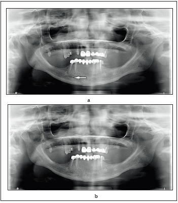
on the panoramic images (Figure 1a) and was accompanied by
an unaltered copy to facilitate identification (Figure 1b). The
responders were asked to report the name of the marked structure
in the space provided, there were no multiple choices.
Figure 1: a. Example of panoramic radiographic image showing region of interest marked by an arrow,
b. Un-altered copy of the same image.
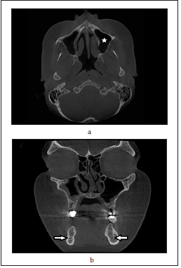
The last section of the survey included four CBCT images of partially edentulous patients that were retrieved from the records of oral and maxillofacial radiology department. Patients records were anonymous with guarantee of data confidentiality. In this section, the participants were asked to choose the type of view from four multiple choices [axial, sagittal, coronal, or panoramic], and to name the marked anatomical structure for each image (Figure 2a & 2b).
Figure 2: Examples of CBCT radiographic images showing regions of interest marked and the question was to
choose the type of view and to identify the anatomical structure marked.
a. axial view, maxillary sinus,
b. coronal view, mandibualr canal.
All responses for the sections three and four were reported
as correct or incorrect, and frequency of correct responses was
reported.
The collected data was analyzed by Statistical Package for
Social Sciences Version 16.0 [IBM SPSS v. 16.0]. The data analysis
was performed according to descriptive statistics and presented
as frequencies [n] and percentages [%]. Students results were
analyzed by Chi-square test to compare responses depending
on level of education and gender. Level of significance was set
at P<0.05.
Result
This study addressed all male and female sixth year students
and dental interns, out of 195 targeted students, 72 responses
were received [response rate 36.9%]. The distribution of students
according to gender and level of education is shown in (Figure 3).
There was significant increase in female responses relative to males
in both levels.
Majority of the participants 95.8% [sixth year students n=46,
interns n=23] knew that panorama means wide view. In response
to the use of panoramic radiography in their routine examination of
patients, 89.6% [n=43] of the sixth-year students and 87.5% [n=21]
interns reported [Yes]. No significant difference between males and
females [P=0.439] or between 6th year and interns [P=0.073]. Only
two female interns denied the use of panoramic radiography in
their routine work.
Majority of participants did not agree that panoramic
radiography is ideal for implant planning. Detailed responses are
shown in (Figure 4).
Figure 3: Pie chart distribution of students according to gender and level of education in the responding sample.
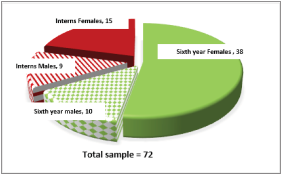
Figure 4:Bar chart students’ response to question: is panoramic radiography ideal in implant planning?

Table 1 shows responses to questions [4-6] of the second part of the questionnaire, concerned with students’ attitude towards CBCT; Have you heard of CBCT used specifically for implant planning? what does CBCT abbreviation stands for? Have you used CBCT in your work before with any of your patients? 95.75% of sixth year students and 87.5% of interns have heard of CBCT used specifically in implant planning. There were no significant differences between males and females or between levels of education.
Table 2:Responses to questions [4-6] concerned with students’ attitude towards CBCT.

Most of the 6th year students 58.3% [n=28], and 50% of interns
[n=12] did not use CBCT with any of their patients before. Mean use
in 6th year students 0.54±0.74, one time is the higher frequency of
use [31.2%]. Mean use in dental interns was 1.16±1.43, where 2-3
times were the higher frequency of use [16.7%] each. Independent
T-test showed significant difference between mean use of sixth
years and dental interns [P= 0.017]. Comparison between males
and females showed that 42.1% [n=8] males and 60.4% [n=32]
females did not use CBCT with their patients, with mean use in
males 1.26 and females 0.56 with statistically significant difference
[P =0.013].
Majority of participants 93.8% [n=45] sixth year students,
and 83.3% [n=20] interns, obtained knowledge of CBCT in their
classes with no significant difference [P=0.173]. other sources of
information revealed were seminars; 22.9% [n=11] sixth year
students and 50% [n=12] interns, with statistically significant
difference [P=0.02]. Followed by information obtained from the
internet; 35.4% [n=17] sixth year students, and 45.85% [n=11]
interns. None of the participants reported any other sources of
knowledge.
Table 2:Participants opinion regarding the use of CBCT in different fields of dentistry.

Table 2 shows participants’ opinion about the use of CBCT in
different dental fields. Highest percent [41.7%] was reported in all
fields of dentistry, followed by maxillofacial surgery [38.9%], and
endodontics [34.7%].
Considerable number of the respondents were willing to receive
extended information and practice in CBCT imaging as shown in
(Figure 5). No significant difference was reported between males
and females [P=0.302], or between sixth year students and interns
[P=0.18].
Figure 5: Bar chart students’ response to question: Are you willing to receive extended information and practice in CBCT imaging.

In response to the questions of section three regarding
identification of anatomical landmarks in panoramic radiography,
high level of correct identifications of the marked anatomical
landmarks was reported for both sixth-year students and dental
interns with average correct score 91.65% and 91.7% respectively
(Figure 6).
However, moderate level of correct identification of cross
section views and marked anatomical landmarks in CBCT
radiography for both sixth-year students and dental interns with
an average correct answers 51.56% each as shown in (Figure 7).
Significant differences in identification of sagittal plane [P=0.021]
was reported.
Figure 6: Bar chart percent of correct identification of anatomical landmarks in panoramic radiography of sixth year students and dental interns.
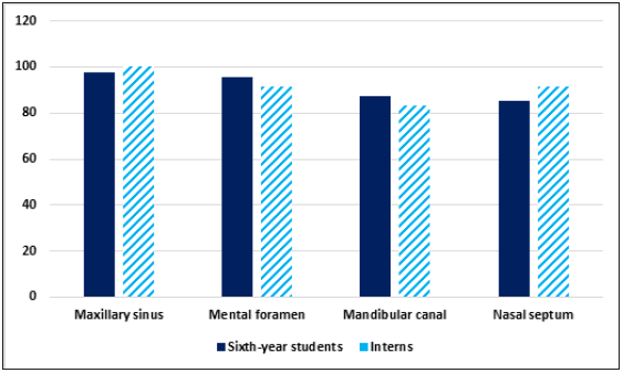
Figure 7: Bar chart percent of correct identification of cross-sectional view and marked anatomical landmarks in CBCT radiography.
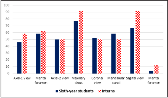
A two-sample t-test of proportions showed significant difference between the proportion of correctly identified anatomical landmarks in panoramic radiography and CBCT of sixth year students [t=9.5, P=0.0005], and dental interns [t=6.724, P=0.0005]. Thus, the null hypothesis was rejected and there was difference between knowledge of panoramic radiography and CBCT.
Discussion
CBCT has been considered a promising technology that is suited
for use in clinical dental practice. CBCT imaging is useful in the
assessment of growth and development. Its value in endodontics,
implant planning, surgical assessment of pathology, TMJ assessment
and pre- and postoperative assessment of craniofacial fractures has
been reported. Although shortcoming may be considered when
using this technology for soft- tissue imaging [7].
An understanding of the anatomical landmarks is necessary
to enhance diagnostic ability. Students’ knowledge regarding
panoramic radiography was assessed in a number of previous
reports [8,9]. Studies assessing students’ knowledge and attitude
regarding CBCT have focused on students’ responses towards
radiographic techniques, applications, and radiation doses [10-
13]. To the authors’ knowledge, there have not been any reports
analysing students’ level of understanding of anatomical findings
in CBCT images. The target sample chosen for this study were the final year dental students and freshly graduated dental interns
who study in their final year curriculum an annual didactic course
on dental implantology. Thus, they are expected to appreciate the
importance of interpretation of anatomical landmarks for implant
treatment planning.
Panoramic radiographs have been used widely as a diagnostic
imaging technique; however, accurate evaluation of hard tissue
morphology and density is difficult because of the variable degree of
distortions, in addition to the lack of information about buccolingual
cross-section dimension or the inclination of the alveolar ridge
[2]. The international congress of oral implantologists 2012,
supported the use of CBCT in dental implant treatment planning
[14]. Accordingly, the aim of the current study was to assess and
compare knowledge and attitude of sixth year dental students and
interns regarding digital panoramic and CBCT radiography.
The study targeted all male and female sixth-year students
and dental interns. A previous study by Al Noaman [11], assessed
the knowledge and attitude of CBCT in undergraduate and post
graduate female dental students only, while this study compared
between responses of both male and female students.
The majority of participants in this study were females,
although that a CBCT facility is available in both male and female
campuses. This reflected the demography of the faculty as a whole
and the direct accessibility of the authors to the participants,
however no differences in most of the responses were found among
female and male students in both levels of education. This high rate
of female’s response compared to males is in concurrence with the
study carried out in Turkey on pre-graduate and post-graduate
dental students in two institutions in Ankara [13].
Knowledge regarding panoramic radiography showed
highest level of correct interpretation. These results reflected the
familiarity of the students to panoramic radiography and agrees
with their admission of the routine use of panoramic radiography
with their patients. These results differ from another study carried
in Japan that reported only 53% rate of correct answers of dental
students in understanding normal panoramic anatomy. This might
be attributed to the difference in the data required in that study
with the need of identification of bone, soft tissue and ghost images
in the radiographs [8]. A different study in Queensland, Australia
reported varying range of correct answers between in revealing
radiographic anatomy, positioning errors, and pathology anomalies
related to panoramic radiographs [9].
Most of the respondents recognise the use of CBCT in implant
planning, however, many of them did not used CBCT with their own
patients before. Higher percentage of the interns reported the use
CBCT. A reasonable finding that reflects that the interns are close
to enter their actual professional careers and are more liberated to
choose their own techniques not restrained by the requirements of
the college curriculum. Despite the moderate rate of utilization of
CBCT by the undergraduates, still they showed higher rate of use
than previous studies in which the students did not have any access
to CBCT unit in their colleges [10,13].
With respect to the sources of information about CBCT as
reported by the participants, the majority acquired much of their
knowledge in faculty classes which exposes the importance of
the curriculum design at this level. The interns reported higher
level of seminars attendance, being graduated renders them more
flexible to attend extra faculty courses and training sessions. These
results differ from Qurashl et al. [12], where minimum percent of
their interns gained CBCT information from attending seminars,
or through using the internet. Current results differed as well from
Noaman [11], who reported that none of the postgraduates attended
any seminars related to CBCT, in contrary to the undergraduates
who attended CBCT-related courses. Kamburoglu et al. [13]
reported that majority of the postgraduates learned about CBCT
in seminars [13]. Dental students at Dow university stated that
textbooks were their main source of information [10].
In the present study, sixth-year students thought that CBCT
would be used most in maxillofacial surgeries and least in
orthodontics. On contrary, most of dental interns supposed that
CBCT would be used in all fields of dentistry, and none of them
thought it would be used in orthodontics. In Mangalore, India, the
interns reported that CBCT would be used in all the fields specified
in the questionnaire, while least suggested its use in orthodontics
[12]. The low choice for orthodontics might be attribute to the
fact that undergraduates study orthodontics basics, a branch that
requires further long postgraduate study to gain deep insights.
The advocated use of CBCT in all fields of dentistry agrees with
the studies at Dow University [10], and in Turkey [13]. Results are
rather different from the previous study carried out by Noaman
[11] where the higher field of CBCT application reported by the
undergraduates was implant dentistry and by the postgraduates
was maxillofacial surgeries and dental implants [11].
Many participants were willing to receive extended information
and practice on CBCT imaging which suggested the awareness
of the new technologies and their applications in different fields
of dentistry and encourages extended learning programs and
workshops. Dental interns in Mangalore, India [12], also suggested
the need of frequent workshops to acquire more knowledge about
CBCT.
In response to the last section of the questionnaire concerned
with identification of the cross-section view and anatomical
landmarks in CBCT radiography, a generally moderate correct
answers were reported for both levels of education which indicated
awareness of the participants of this type of radiology but who
are in need of more practice and training for better diagnosis
and treatment planning. It was noticed that the highest correct
identification was of the maxillary sinus in the axial view. The
lowest was the mental foramen in the sagittal view where most of
the answers were “I don’t know” and to a lesser extend confused
with mandibular canal. The fact that the participants did not have
access to the whole volume of the CBCT radiography might be one
of the reasons contributing to the misjudgment, in addition to the
minimum use of CBCT reported.
These results indicate that dental curriculum designers should consider the increased CBCT practice and training in the undergraduate levels, side by side with the theoretical knowledge and to provide education focused on landmarks with lower rates for correct answers.
Conclusion
CBCT is one of the most significant developments in modern dentistry, which should be properly understood by dentists and dental students. This study was carried out in a dental institution that has a CBCT facility, yet the present study showed that final year dental students and interns were more used to panoramic radiography than CBCT. Students showed moderate awareness of CBCT implant-relative anatomy which suggests that more knowledge of CBCT should be gained through regular continuing education programmes, post graduate education courses, and meetings and seminars to update dentists’ knowledge and improve their understanding of radiographic images.
Appendix 1
Part [1]: Personal information
1. l- Gender* Male [ ] Female. [ ]
2. University of graduation* …………….
3. 2- Year of graduation* ………………….
Part [2]: General information and attitude toward Panoramic and CBCT radiographs
1. What does panoramic mean?*
Narrow view [ ] Wide view. [ ]
Focused view [ ] Other: [ ]
2. Do you use Panoramic Radiograph in your routine examination of patients? *
Yes [ ] No [ ] Maybe [ ]
3. Do you think panoramic radiographs are ideal for implant planning?
Yes [ ] No [ ]
4. Have you heard of CBCT used specifically for Implant planning? *
Yes [ ] No [ ]
5. What does CBCT abbreviation stands for? *
………………………………………………………………
6. Have you used CBCT in your work before with any of your patients? *
Yes [ ] No [ ]
7. If YES to previous question, how many times? ………
8. How do you obtain information regarding CBCT? * [Multiple responses are allowed.]
Faculty lessons [ ]. Seminars [ ]. Internet [ ]
Others. [ ] Please specify …………………………………
9. To what extent do you think CBCT will be used in routine dental practice in the near future? *
[Multiple responses are allowed.]
In all areas of dentistry [ ]
Prosthodontics. [ ]
Endodontics. [ ]
Oral and maxillofacial surgery [ ]
Orthodontics [ ]
It will not be commonly used in routine practice [ ]
No idea [ ]
10. Are you willing to receive extended information and practice in CBCT imaging? *
Yes [ ] No [ ]
Maybe [ ] No idea [ ]
Part [3]: Identifying anatomical landmarks related to implant surgery in Orthopantomogram [OPG]
You will find orthopatogram images of partially edentulous patients retrieved from the records of Oral and Maxillofacial Radiology
Department. If you, please identify marked anatomical structures in each image. You will find the same image twice, the top one with a
mark on the identifiable structure, the bottom one without any marks for clarification.
Identify structures marked:
(1) Case 1*:
(2) Case 2*:
(3) Case 3*:
(4) Case 4*:
Part [4]: Identifying anatomical landmark related to implant surgery in CBCT
These are CBCT images of partially edentulous patients retrieved from the from the records of Oral and Maxillofacial Radiology Department.
If you, please choose the type of view in each image and identify the marked anatomical structure.
Type of View: *
Axial [ ]
Coronal [ ]
Lateral. [ ]
Panoramic [ ]
No Idea. [ ]
Marked Anatomical structure:*
References
- Elani HW, Starr JR, Da Silva JD, Gallucci GO (2018) Trends in dental implant use in the U.S., 1999-2016, and projections to 2026. J Dent Res 97(13): 1424-1430.
- Bagchi P, Nikhil J (2012) Role of radiographic evaluation in treatment planning for dental implants: A review. J Dent Allied Sci 1(1): 21-25.
- Athota A, Gandhi Babu DB, Nagalaxmi V, Raghoji S, Waghray S, et al. (2017) A comparative study of digital radiography, panoramic radiography, and computed tomography in dental implant procedures. J Indian Acad Oral Med Radiol 29(2): 106-110.
- Shahidi S ZB, Abolvardi M, Akhlaghian M, Paknahad M (2018) Comparison of dental panoramic radiography and cbct for measuring vertical bone height in different horizontal locations of posterior mandibular alveolar process. J Dent (Shiraz) 19(2): 83-91.
- Bathwarr N, Nahar P (2015) Diagnostic imaging in implant dentistry: A review. J Pacif Acad High Edu Res 6(4): 32-40.
- Ozalp O, Tezerisener HA, Kocabalkan B, Büyükkaplan UŞ, Özarslan MM, et al. (2018) Comparing the precision of panoramic radiography and cone-beam computed tomography in avoiding anatomical structures critical to dental implant surgery: A retrospective study. Imaging Sci Dent 48(4): 269-275.
- William CS, Farman AG, Predag S (2006) Clinical applications of cone-beam computed tomography in dental practice. J Can Dent Assoc 72(1): 75-80.
- Maeda N, Hosoki H, Yoshida M, Suito H, Honda E (2018) Dental students’ levels of understanding normal panoramic anatomy. J Dent Sci 13(4): 374-377.
- McNab S, Monsour P, Madden D, Gannaway D (2015) Knowledge of undergraduate and graduate dentists and dental therapists concerning panoramic radiographs: knowledge of panoramic radiographs. J Dent Oral Med 3(2): 46-52.
- Iqbal W, Farooq F, Abdulbari Y, Nazir F, Quadri MA, et al. (2016) Knowledge, attitude and practice regarding computed tomography and cone beam computed tomography among dental students at dow university of health sciences. Adv Dent Oral Health 2(4): 001-0016.
- Noaman R, Khateeb S (2017) Knowledge and attitude of cone beam CT- a questionnaire-based study among Saudi dental students. Bri J Med Med Res 19(4): 1-10.
- Qurashl N, Chatra L, Shenoy P, Veena KM, Prabhu R (2018) Knowledge and attitude about cone beam computed tomography [CBCT] among dental interns. J Dent Res 64: 19-25.
- Kamburoglu K, Kursun S, Akarslan ZZ (2011) Dental students' knowledge and attitudes towards cone beam computed tomography in Turkey. Dentomaxillofac Radiol 40(7): 439-443.
- Benavides E, Rios HF, Ganz SD, An CH, Resnik R, et al. (2012) Use of cone beam computed tomography in implant dentistry: The International Congress of Oral Implantologists consensus report. Implant Dent 21(2): 78-86.
© 2021 Rania Mostafa Moussa. This is an open access article distributed under the terms of the Creative Commons Attribution License , which permits unrestricted use, distribution, and build upon your work non-commercially.
 a Creative Commons Attribution 4.0 International License. Based on a work at www.crimsonpublishers.com.
Best viewed in


.jpg)


 Editorial Board Registrations
 Submit your Article
 Refer a Friend
 Advertise With Us
.jpg)


.jpg)


.bmp)
.jpg)
.png)
.jpg)


.jpg)


.png)

.png)


.png)


