- Submissions

Full Text
Modern Research in Dentistry
Prosthodontic Treatment for Edentulous Patient with Bell’s Palsy: A Case Report
Alvydas Gleiznys1* and Zivile Zidonyte2
1Department of Prosthodontics, Faculty of Odontology, Medical Academy, Lithuanian University of Health Sciences, Lithuania
2Faculty of Odontology, Medical Academy, Lithuanian University of Health Sciences, Lithuania
*Corresponding author: Alvydas Gleiznys, Department of Prosthodontics, Faculty of Odontology, Medical Academy, Lithuanian University of Health Sciences, Lithuania
Submission: March 19, 2020;Published: May 29, 2020

ISSN:2637-7764Volume5 Issue2
Abstract
Bell’s palsy is an idiopathic neuropathy of the facial nerve, meaning that a cause is unknown. It is usually recognized as an acute weakness and disability to move one side of the entire face. Problems of speech, swallowing and eating occur and the chance of the quick recovery takes 6 months (85% of patients) or is impossible at all. However, it is inevitable to restore patient’s masticatory system and to return the ability to live a comprehensive life. The purpose of the work is to report a case of Bell’s Palsy, reveal advices and difficulties that were met during this treatment.
Methods: A clinical and radiographic examination for the 51-year-old patient was made in accordance with neurologist, otorhinolaryngologist and general doctor. Accordingly, an electronic research was performed on databases such as The Cochrane Library, EMBASE via Science Direct, MEDLINE via PubMed in order to collect as much information as possible. All selected data was summarized and the protocol of treatment for a patient was determined.
Result: A clinical case presents our findings and the protocol of the treatment, replenished with data from scientific literature. The main aspects are mentioned with our plans to continue our research how to improve prosthodontic treatment for patients with facial paralysis.
Conclusion: Clinical and radiographic analysis has showed a need for a specific treatment. According to the clinical case, the main anatomical structures were marked according which a required border molding has to be reached for all types of patients and have distinguished the ones that are inherent for patients with Bell’s palsy.
Keywords: Bell’s palsy; Complete dentures; Border molding; Custom trays
Introduction
Bell’s palsy is known as facial paralysis that has an idiopathic cause. This severe condition can appear because of unknown conditions and complicates one’s life [1].
The main features of the Bell’s palsy are:
- Facial droop and difficulty making facial expressions, such as closing eye or smiling;
- Increased sensitivity of sound on the paralyzed side;
- Pain through the jaw on the affected side;
- A decreased ability to eat, chew, talk [1-3].
Many different treatment approaches have been offered by neurologist to the resolution for such one side paralysis and a patient was motivated to accept dental treatment to recover his masticatory system. In prosthodontics it appears indispensable to transfer the view from the mouth to the casts. Sometimes mistakes occur and collaboration between dental technician and a dentist interrupts, wherefore appropriate treatment cannot be accomplished [4-9]. Accordingly, one of the most important factors how to maintain this professional communication is taking into account the correct determination of custom tray borders, border molding and impressions [4-8]. Treatment becomes especially difficult in edentulous patients with health disorders, so that we have decided to announce our clinical case. The purpose of this case report study was to perform an objective treatment for a patient with one side facial paralysis, to determine the borders of an upper jaw custom tray and to assure impression is ready to be sent to the laboratory [7,9,10].
Case Report
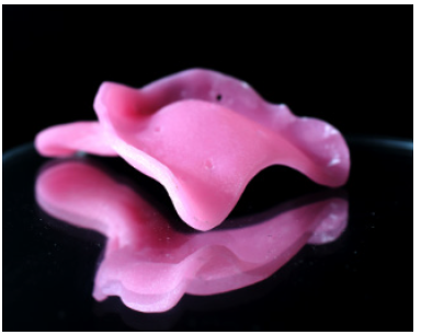
The case presented is of a 51-year-old male patient who came to our Clinic with a medical history of Bell’s palsy. Treatment plan was based on electronic research and the experience our doctors. The research was based on three databases (The Cochrane Library, EMBASE via Science Direct, MEDLINE via PubMed). It was performed by using keywords: “complete dentures”, “border molding”, “and custom trays”, “ Bell ’s palsy”. As inclusion criteria we have chosen clinical cases on humans, English language, articles that have proven statistically confirmed value. After screening the literature, detailed information was used for the treatment. It was decided to extract teeth that cannot be restored. After 4 weeks, preliminary impressions were made with alginate and poured in Type III dental stone (Figure 1). After fabricating primary casts, custom acrylic resin trays were made and single-step border molding was done (Figure 2).
Figure 1: Primary casts.
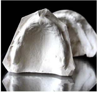
Figure 2: Upper jaw custom tray.
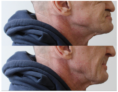
By customizing individual trays and taking trial impressions, a few main structures have shown a need to be checked whether custom tray border molding is correct. After corrections and single-step border molding, a final impression was taken. Accordingly, an asymmetry of polyvinyl siloxane impression between both sides has shown manifesting differences between normal and paralyzed sides (Figure 3). From the photo it is clear that a deviation of the labial frenum is linked to the paralyzed side, right buccal frenum is not expressed enough and the right labial and buccal sulcus take less space than the left ones. However, other structures are similar in both sides (Figure 4). On the active side we could make functional moves in order to show off muscle and mucous membrane activity, whilst the passive one only showed us a regular anatomy without expressed amplitude of intraoral structures and soft tissues. Even though a patient was incapable of moving the right side of his face, during functional impressions his right side of the case was manipulated by a doctor in order to achieve the best retention and stability. Further treatment protocol steps were made the same as for conventional complete dentures. Denture bases and wax rims were made to record maxilla mandibular relationship. VDO (vertical dimension of occlusion) was determined to be 2-3mm less than VDR (vertical dimension of rest). Accordingly, the goal was successfully accomplished-dentures have shown a satisfaction with the chewing efficiency. Patient’s articulating, ability to talk and better esthetic appearance were improved as well (Figure 5&6).
Figure 3: Impression.
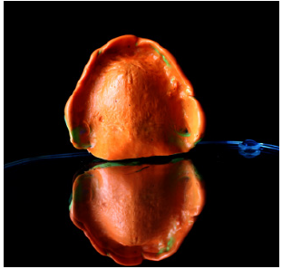
Figure 4: Anatomical landmarks.
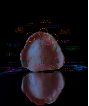
Figure 5: Face frontal figure.

Figure 6: Face profile figure.
Conclusion
One side facial paralysis has always been a challenge not only for neurologists, otorhinolaryngologist and general doctors; it creates the same difficulties for restorative dentistry specialists. This case report presents an inexpensive and simple technique how to evaluate whether an upper jaw impression is finished and can be used for dentures fabrication. After analyzing custom trays, functional impressions and casts, the main anatomical structures that have to be reviewed carefully were sorted out: the labial frenum is linked to the paralyzed side, right buccal frenum is not expressed and the right labial and buccal sulcus take less space than the left ones. Finally, a patient has shown a satisfaction and can spend his days more comfortable and live comprehensive life.
References
- Hauser W, Karnes W, Annis J, Kurland L (1971) Incidence and prognosis of Bell’s palsy in the population of Rochester, Minnesota. Mayo Clin Proc 46(4): 258-264.
- Yanagihara N (1988) Incidence of Bell’s palsy. Ann Otol Rhinol Laryngol 97: 3-4.
- Katusic SK, Beard CM, Wiederholt WC, Bergstralh EL, Kurland LT (1986) Incidence, clinical features, and prognosis in Bell’s palsy, Rochester, Minnesota, 1968-1982. Ann Neurol 20(5): 622- 627.
- Komagamine Y, Kanazawa M, Sato Y, Iwaki M, Jo A, Minakuchi S, et al. (2019) Masticatory performance of different impression methods for complete denture fabrication: A randomized controlled trial. Journal of Dentistry 83: 7-11.
- Namratha N, Shetty V (2014) A technique to evaluate custom tray border extensions before peripheral molding. The Journal of Prosthetic Dentistry 112(6): 1603-1604.
- Kaur S, Datta K, Gupta S, Suman N (2016) Comparative analysis of the retention of maxillary denture base with and without border molding using zinc oxide eugenol impression paste. Indian Journal of Dentistry 7(1): 1-5.
- Shopova D, Slavchev D (2019) Laboratory investigation of accuracy of impression materials for border molding. Folia Medica 61(3): 435-443.
- Pawar R, Kulkarni R, Raipure P (2018) A modified technique for single-step border molding. The Journal of Prosthetic Dentistry 120(5): 654-657.
- Lo Russo L, Caradonna G, Troiano G, Salamini A, Guida L, et al. (2020) Three-dimensional differences between intraoral scans and conventional impressions of edentulous jaws: A clinical study. The Journal of Prosthetic Dentistry 123(2): 264-268.
- Munakata Y, Kasai S (2007) Determination of occlusal vertical dimension by means of controlled pressure against tissues supporting a complete denture. J Oral Rehabil 17(2): 145-150.
© 2020 Alvydas Gleiznys. This is an open access article distributed under the terms of the Creative Commons Attribution License , which permits unrestricted use, distribution, and build upon your work non-commercially.
 a Creative Commons Attribution 4.0 International License. Based on a work at www.crimsonpublishers.com.
Best viewed in


.jpg)


 Editorial Board Registrations
 Submit your Article
 Refer a Friend
 Advertise With Us
.jpg)


.jpg)


.bmp)
.jpg)
.png)
.jpg)


.jpg)


.png)

.png)


.png)


