- Submissions

Full Text
Modern Research in Dentistry
Horizontal Ridge Augmentation with the Cortical Lamina Technique: A Case Report
Roberto Rossi2, Alessandro Conti2, Davide Bertazzo3 and Andrea PILLONI*1
1University of Roma Sapienza, Italy
2Private Practice, Genova, Italy
3CDT Casale Monferrato, Italy
*Corresponding author: Andrea Pilloni, Professor and Chairman of Periodontology, University of Roma ‘Sapienza’, Roma, Italy
Submission: October 29, 2019;Published: November 12, 2019

ISSN:2637-7764Volume4 Issue4
Abstract
Ridge defects are a common problem following tooth extraction, the literature shows that horizontal defects seem to be the easier to correct. Nevertheless a number of procedures, matherials and methods have been proposed to treat such problem and the use of a combination of a xenograft associated with some autogenous bone and resorbable membrane seems to be the best predictable option. The cortical lamina technique has been introduced about ten years ago and both clinica and hystological studies have validated its use. In this case presentation we will show how the cortical lamina, significantly, changed the anatomy of a severely resorbed mandible in order to accommodate and restore dental implants.
Introduction
Tooth loss is a common unfortunate event in dentistry, quite often following an extraction we observe a certain degree of remodeling in both the hard and soft tissue [1]. Ridge defects can also be caused by faulty extractions, traumatisms, periodontal disease and peri-implantitis. These defects can show different patterns, some with an horizontal pattern, some with a vertical component and some showing a combination of both horizontal and vertical hard and soft tissue loss. Many different surgical approaches have been offered for the resolution of such problem, and a recent systematic review done by Sanz-Sanches et al. 2015 indicated that the most reliable solution to solve horizontal defects is the combination of a xenograft associated with a resorbable membrane [2]. A new technique that is showing promising result in horizontal ridge augmentation is the cortical lamina and uses a membrane made of cortical porcine bone [3,4], several publications and reports prove its usefulness and efficacy in treating ridge defects. The Cortical lamina technique is becoming a well understood and used procedure for both horizintal and vertical bone augmentation [5,6]. It is basically a semi rigid, flexible, collagenated xenogenic bone membrane (Tecnoss, Coazze, TO, Italy) that has the advantage to integrate along with the bone graft placed underneath and to fully integrate in the area wher it is placed, reshaping and augmenting the area [5].
Case Presentation
The case presented is of a female patient 41 years old with a failing long span bridge in
the lower left quadrant. Her CBCt (Figure 1&2) shows a severe pattern of bone resorbtion, the
ridge is basically knife edged and poorly represented in its cancellous component. Clinically
also the soft tissue show (Figure 3) this pattern with a high insertion of the vestibule. The
patient had a negative medical history and there was no contra-indication for GBR and
subsequent implant treatment. After local anesthesia with Articaine 1:200.000 full thickness
buccal and lingual flaps were elevated to expose the very narrow edentulous ridge (Figure
4). With the aid of a sharp piezo-surgical tip the narrow ridge was perforated in the buccal
aspect to promote some bleeding from the marrow spaces and in order to hydrate the bone
graft placed to its buccal aspect. It was a mix of autogenous bone scarped from the area and
collagenated porcine bone (GenOs, Tecnoss, Coazze, TO, Italy), the newly shaped ridge (Figure
5) was than covered with a curved cortical lamina, cut and shaped in order to fit the area and
protect the underlying grafted ridge (Figure 6).
The falps were than advanced to accommodate the new volume and sutured with PTFE 4.0
sutures. 8 Months after surgery one can notice the new aspect of the ridge (Figure 7) and after flaps were opened to exposed the new ridge one can notice that the
new horizontal volume coud than receive 4.2mm diameter implants
(Resista, Omegna, Italy) without any problem (Figure 8). The next
picture (Figure 9) shows how the two fixtures were properly
adjusted to the new anatomy. Three monts later the implants were
exposed, healing abutments were connected to further condition
and widen the band of keratinized gingiva by apically repositioned
flap (Figure 10). In the next two pictures (Figure 11&12) we can
see the final restoration, a screwed in monolithic zirconia two unit
bridge, that fits well the previously edentulous ridge and offers a
very natural and bio-mimetic emergence profile. At last (Figure
13-16) the radiographic history of the case, with the edentulous
ridge and the old long span bridge, the implants placed after ridge
augmentation, its relationship to the bone crest at the time of
impressions and four years after the final restoration was delivered.
Figure 1: CBCT.
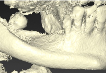
Figure 2: Slice of CBCT.
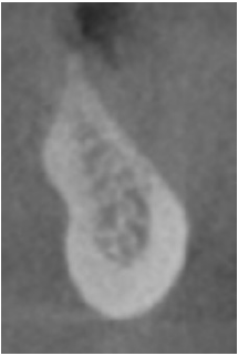
Figure 3: Appearance of the edentulous ridge.

Figure 4: Extremely resorbed ridge.
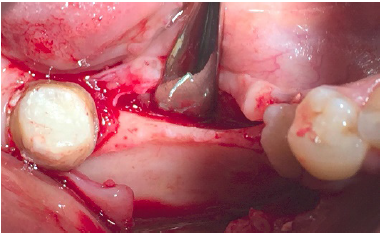
Figure 5: Aspect of the grafted ridge.
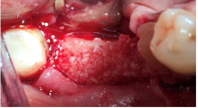
Figure 6: application of cortical lamina.
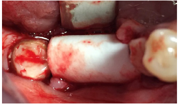
Figure 7: Healing at 8 months post op.
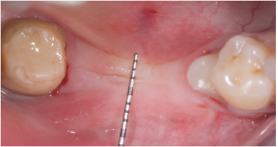
Figure 8:Insertion of two 4.2mm fixtures.
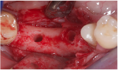
Figure 9: implants in place.
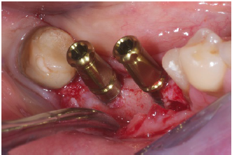
Figure 10:Healing abutments.
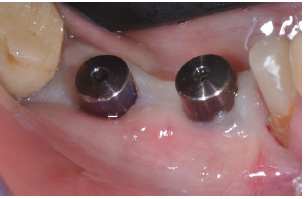
Figure 11:Final restoration in place.
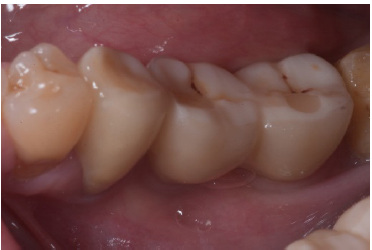
Figure 12:proper emergence profile.
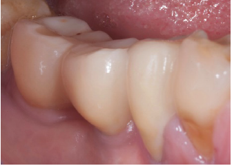
Figure 13:Initial rx.
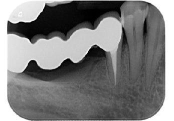
Figure 14:rx implants.
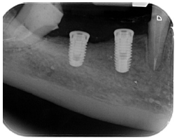
Figure 15:Impression.
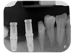
Figure 16:Final restoration (4y).

Conclusion
Ridge defects have always been a challenge for the clinician and many surgical options and bio-materials have been advocvated for its treatment. In recent years the cortical lamina technique has surfaced as one of the techniques that reliably works in the solution of such problem. The case presented in this article had at baseline an horizontal ridge defect that would have not suggested the use of dental implants. Following ridge augmentation with a Xenograft covered with the cortical lamina it was possible to insert standard diameter implants and later to restore them. An important point is that even after four years of occlusal loading the regenerated bone does not show any kind of remodeling and/or resorbtion, thus confirming the efficacy of this treatment modality for horizontal ridge defects.
References
- Schropp L, Wenzel A, Kostopoulos L, Karring T (2003) Bone healing and soft tissue contour changes following single-tooth extraction: a clinicala and radiographic 12-months prospective study. Int J Periodntics Restorative Dent 23(4): 313-323.
- Sanz Sanchez I, Ortiz Vigon, Sanz Martin I, Figuero E, Sanz M (2015) Effectiveness of lateral bone augmentation on the alveolar crest dimension-a systematic review and meta-analysis. J Dent Res 94(9 Suppl): 128S-142S.
- Wachtel H, Helf C, Thalmair T (2014) The bone lamina technique: a novel approach to bone augmentation. Bone Biomaterials and Beyond, EDRA Lswr.
- Rossi R, Foce E, Scolavino S (2017) The cortical lamina technique a new option for ridge augmentation procedure: protocol and pilot case. J Leb Dent Ass 52(1).
- Rossi R, Rancitelli D, Poli PP, Rasia da Polo M, Nannmark U, et al. (2016) The use of collagenated porcine cortical lamina in the reconstruction of alveolar ridge defects: a clinical and histological study. Minerva Stomatol 65(5): 257-268.
- Pagliani L, Andersson P, Lanza M, Nappo A, Verrocchi D, et al. (2012) A collagenated porcine bone substitute for augmentation at Neoss implant sites: a prospective 1-year multicenter case series with histology. Clin Impl Dent Relat Res 14(5): 746-758.
© 2019 Roberto Rossi. This is an open access article distributed under the terms of the Creative Commons Attribution License , which permits unrestricted use, distribution, and build upon your work non-commercially.
 a Creative Commons Attribution 4.0 International License. Based on a work at www.crimsonpublishers.com.
Best viewed in


.jpg)


 Editorial Board Registrations
 Submit your Article
 Refer a Friend
 Advertise With Us
.jpg)


.jpg)


.bmp)
.jpg)
.png)
.jpg)


.jpg)


.png)

.png)


.png)


