- Submissions

Full Text
Modern Research in Dentistry
The Marvel of 3D Imaging: A Case of an Undetectable Condylar Split Fracture
Geon Pauly1* and Navami Ashok2
1 Independent Researcher, India
2 Department of Public Health Dentistry, Vydehi Institute of Dental Sciences and Research, India
*Corresponding author:Geon Pauly, Independent Researcher, Bhatar Road, Surat, PIN-395017, Gujarat, India, Tel: +918905102696, Email: geonpauly@gmail.com
Submission: August 06, 2018;;Published: October 25, 2018

ISSN:2637-7764Volume3 Issue3
Abstract
Radiography has become one of the cornerstones for palatable diagnosis in the world of medical sciences. Even amongst our dental fraternity; the use of conventional radiographs in day-to-day cases has increased considerably over the past few decades. With the arrival of 3D imaging, the credibility of oral diagnosis has leapfrogged to a different level. The purpose of this article is to present a case of an undetectable split fracture of the condyle, only made perceptible, courtesy 3D imaging.
keywords: Three-dimensional imaging; Bone fractures; Radiography; Oral diagnosis
Introduction
Imaging is one of the most important tools for dentists worldwide to not only diagnose difficult cases but also to design an effective treatment plan. Dentists routinely use 2-dimensional (2D) static imaging techniques, whether analogue or digital. Despite the imaging modality like orthopantomograph (OPG) which has become the global standard as a screening diagnostic tool; in some cases, findings such as the deepness of structures, the finer details of a positive finding or the extent of the pathosis cannot be obtained and localized with 2D imaging. Three-dimensional (3D) imaging was developed in the early of 1990’s and has gained a precious place in dentistry ever since. The advent of cone beam computed tomography (CBCT) has opened new horizons in the field of diagnostic imaging [1]. Hereby, we discuss a case of a condylar split fracture which was otherwise undetectable even with the aid of conventional radiography.
Case Report
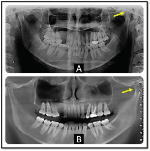
Figure 1:No Visible Fracture Seen On: A-An OPG Image, B-A Panoramic Image of CBCT.

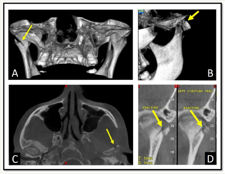
A 29-year-old female patient reported to our facility with a chief complaint of intermittent pain on the left side of the face since, one month. She gave a history of a fall one month prior. Past medical, dental and personal histories were non-contributory. Extra-oral examination revealed pain localized to the pre-auricular region on the left side and was tender on palpation. An OPG image revealed no abnormal findings (Figure 1). CBCT images revealed a distinct split fracture on the left condylar head extending about one cm in length which was undetectable otherwise (Figure 2). The patient was further referred to an oral and maxillofacial surgery for the needful.
Figure 2:CBCT Images Showing Split Fracture of Left Condyle On: A-3D Image: Rear View, B-3D Image: Left Lateral View, C-Axial Slice, D-Cross-Sectional Slices.

Discussion
Facial fractures may occur in isolation or accompanied by tissue injuries. They may be symptomatic or asymptomatic. However, without timely diagnosis and adequate and appropriate treatment; serious functional, and esthetical problems may emerge [2,3]. Boeddinghaus and Whyte noted that while standard 2D radiographs may help in preliminary examination of maxillofacial trauma, diagnosis of an occult fracture may require tomographic imaging techniques, such as CBCT, that can provide excellent imaging of bony structures [4]. In line with this assertion, the occult fracture diagnosed by CBCT in the present case could not be distinguished by panoramic radiography. Maxillofacial trauma causes severe clinical problems because of the anatomical characteristics of the region and 34% of the cases are accompanied by major trauma [5]. 3D imaging is thus essential for locating anatomic and pathologic components and can provide views of both hard and soft tissue, whereas 2D projections are of limited use because of superimposition, magnification, distortion and misrepresentation of structures [6]. However, the emerging technique CBCT cannot be taken as the primary choice of imaging maxillofacial trauma due to lack of availability and increased radiation exposure. Nevertheless, when weighing the advantages this imaging procedure could be the modality of choice depending on the case. Thus, CBCT seems to be a good complementary and not necessarily a substitute for panoramic radiography in trauma cases.
References
- Karatas OH, Toy E (2014) Three-Dimensional Imaging Techniques: A Literature Review. Eur J Dent 8(1): 132-140.
- Simonds JS, Whitlow CT, Chen YM, Williams DW (2011) Isolated Fractures of the Posterior Maxillary Sinus: CT Appearance and Proposed Mechanism. American Journal of Neuroradiology 32(3): 468-470.
- Açıkel C, Saray A, Yılmaz KB (2011) The Case Report of Maxillofacial Trauma That Required Reoperation. ACU SağlıkBilDerg 2: 49-51.
- Boeddinghaus R, Whyte A (2008) Current Concepts in Maxillofacial Imaging. European Journal of Radiology 66(3): 396-418.
- Malara P, Malara B, Drugacz J (2006) Characteristics of Maxillofacial Injuries Resulting from Road Traffic Accidents-A 5 Year Review of the Case Records from Department of Maxillofacial Surgery in Katowice, Poland. Head Face Med 2: 27-34.
- Scarfe WC (2005) Imaging of Maxillofacial Trauma: Evolutions and Emerging Revolutions. Oral Surg Oral Med Oral Pathol Oral Radiol Endod 100: 75-96.
© 2018 Geon Pauly. This is an open access article distributed under the terms of the Creative Commons Attribution License , which permits unrestricted use, distribution, and build upon your work non-commercially.
 a Creative Commons Attribution 4.0 International License. Based on a work at www.crimsonpublishers.com.
Best viewed in


.jpg)


 Editorial Board Registrations
 Submit your Article
 Refer a Friend
 Advertise With Us
.jpg)


.jpg)


.bmp)
.jpg)
.png)
.jpg)


.jpg)


.png)

.png)


.png)


