- Submissions

Full Text
Modern Research in Dentistry
Implant-Supported Fixed Partial Denture in a Patient with Seckel Syndrome
Arwa AlSayed*
College of Dentistry, Dar Al-Uloom University, Riyadh, Saudi Arabia
*Corresponding author: Arwa ALSayed BDS, MS, MSc, MCD, College of Dentistry, Dar Al-Uloom University, Po Box 2993, Riyadh 11461, Saudi Arabia
Submission: April 09, 2018;Published: April 17, 2018

ISSN:2637-7764Volume2 Issue2
Summary
Seckel syndrome or Microcephalic primordial dwarfism is a rare, autosomal recessive, congenital disorder characterized by severe intrauterine and postnatal growth retardation, microcephaly, mental retardation, and typical facial appearance with beaklike protrusion of the midface. Dental abnormalities include enamel hypoplasia, hypodontia, microdontia, taurodontic root morphology and a high-arched palate. This is a case report of implant supported dental prosthesis in a 19-year-old male with Seckel syndrome. Two implant were placed in the area of #12 and 22. Three months’ post implantation, the site was examined to ensure the osseointegration. A four-unit implant supported screw-retained porcelain fused to metal bridge was attached. Occlusion was checked and oral hygiene instructions were given to the mother. The case was followed up for three years. The osseointegration was excellent with no untoward findings. To the best of our knowledge, this is the first report of implant supported fixed partial denture in Seckel Syndrome patient.
Introduction
Seckel syndrome (SS) is an extremely rare disease that is a form of primordial autosomal recessive dwarfism (MIM 210600). Etiology of the syndrome involves multiple malformations in which both sexes are equally affected [1]. Seckel [2] first described the syndrome in 1960 based on 13 cases from the literature and two personally studied cases. He defines the syndrome as severe intrauterine growth retardation, severe short stature, severe microcephaly, bird-headed profile, receding chin and forehead, large beaked nose, mental retardation, and other congenital anomalies [2].
More than 100 cases have been reported, and it occurs in 1 in 10,000 children without sex preference. Characteristic skeletal anomalies include premature closure of the cranial sutures and fifth finger clinodactyly. In addition to the characteristic craniofacial dysmorphism and skeletal defects, abnormalities have been described in the cardiovascular, hematopoietic, endocrine, and central nervous systems. Retarded bone age and moderate to-severe mental retardation are observed in patients with SS. Other systemic manifestations associated with SS include Fanconi anemia, leukemia, chronic nephritis, dysgenesis of cerebral cortex, and corpus callosum, as well as a vast spectrum of skeletal defects (Sisodia et al 2018).
The phenotypic appearance of the head is well described. The dentition has been described sporadically, focusing on absence of teeth and atrophy or hypoplasia of the enamel (Gellis et al., 1967, Sauk et al 1973). Tooth malformations have often been associated with the syndrome (Muller et al., 1978). Agenesis of two or more teeth has been reported in four Iraqi children, and all children lacked the upper lateral incisors. Short roots and taurodontic molars were consistent characteristics. Sex differences in the dentition were evident. Profound, untreated caries was diagnosed in several deciduous and permanent teeth [3].
Dental malformations in Seckel Syndrome include hypodontia, enamel hypoplasia, crowding, and Class II malocclusion. Amy et al. [4] also reported gingival hyperplasia, recession and ulceration, significant crowding, and early exfoliation of the primary dentition with accelerated eruption of the permanent dentition in girl patient. The Seckel syndrome is characterized by more severe intellectual disability as well as more often the presence of characteristic facial features [5,6].
Case Report
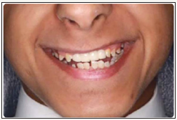
A 19-year-old Saudi male patient diagnosed with a Seckel syndrome reported to the clinic for replacement of upper anterior teeth #12, #11, #21, #22 (Figure 1). He was born to healthy, consanguineous (first cousins) parents. Patient’s mother reported that he was suffering psychologically because of his appearance and his inability to speak properly. Radiographic examination revealed that the remaining permanent teeth have erupted with minimal root development (Figure 2). Periodontal examination showed generalized moderate marginal gingivitis (Plaque score 35%, Bleeding score 15%). Oral hygiene regimen was given to the mother and full mouth gross scaling was performed. Informed consent was obtained for restoring missing teeth with dental implants. Medical clearance was achieved and implants were placed under nitrous oxide sedation and local anesthesia. A variety of tracheal tubes were at hand as many of these young patients have smaller trachea than would be suspected by age and physical size. A full mucoperiosteal flap was raised (Figure 3) and two implants (3.3x8BL- SLActive (Straumann –ITI) were placed in the area of #12 and 22 (Figure 4). Implant sites were grafted buccaly with bone graft (Puros Allograft –Cortico cancellous, Tutogen, Zimmer Dental) and covered with resorbable membrane (Copios pericardium – RTI Biologics, Tutogen, Zimmer Dental). Flap was repositioned and sutured with 4-0 resorbable Vicryl suture (Figure 5). Peri-apical radiograph was taken to show the position of the 2 implants in the edentulous sites (Figure 6). Three months post implantation period, the site was examined for bone and soft tissue healing around the implants to ensure osseointegration. A four-unit implant supported screwretained porcelain fused to metal bridge was attached (Figure 7) and peri-apical radiograph was taken to ensure complete and accurate fitting of the bridge (Figure 8). Occlusion was checked and oral hygiene instructions were given to the mother. The case was followed up for three years (Figure 9). The osseointegration was excellent with no untoward findings. Also, upon follow up, it was noted that the patient was considerably happier and the mother reported that the procedure has changed her son’s life. To the best of the author knowledge, this is the first report of implant supported fixed partial denture in Seckel Syndrome patient [4,7-10].
Figure 1: Frontal view showing the edentulous area with missing teeth #12, 11, 21, 22.
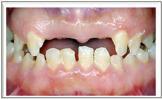
Figure 2: Peri-apical radiograph shows the amount of available bone at the edentulous area (~10mm height).
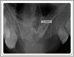
Figure 3: Full mucoperiosteal flap shows a thin bone crest (~4mm).

Figure 4: Narrow BL Straumann implants (3.3 X 8) were placed at #12, 22.
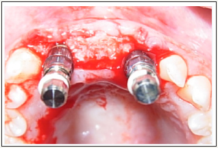
Figure 5: Bone graft and membrane were placed at the buccal ridge to augment the implant sites. Flap was sutured around the implant’s healing abutments.
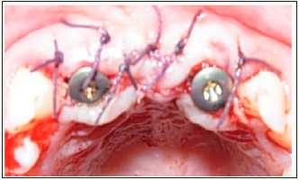
Figure 6: Peri-apical radiograph showing the position of the 2 implants.
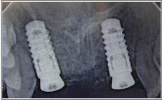
Figure 7: Insertion of a screw retained 4-units PFM bridge supported by the 2 implants at #12, 22
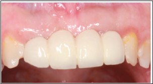
Figure 8: Insertion of a screw retained 4-units PFM bridge supported by the 2 implants at #12, 22
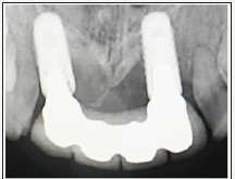
Figure 9: Insertion of a screw retained 4-units PFM bridge supported by the 2 implants at #12, 22
Conclusion
Based on this report, it can be concluded that dental implants can be used in rehabilitation of edentulous areas in Seckel Syndrome patients. Further trials with longer period of follow up are recommended to ensure the feasibility of this treatment for this patient population.
References
- Shanske A, Caride DG, Menasse-Palmer L, Bogdanow A, Marion RW (1997) Central nervous system anomalies in Seckel Syndrome: Report of a new family and review of the literature. Am J Med Genet 70(2): 155- 158.
- Seckel HP (1960) Studies in developmental anthropology including human proportions. S Karger (Ed.), Springfield, C.C. Thomas: Basel-New York, USA, pp. 111.
- Kjaer I, Hansen N, Becktor KB, Birkebaek N, Balslev T (2001) Craniofacial morphology, dentition, and skeletal maturity in four siblings with Seckel syndrome. Cleft Palate Craniofac J 38(6): 645-651.
- Regen A, Nelson LP, Woo SB (2010) Dental manifestations associated with seckel syndrome type II: a case report. Pediatr Dent 32(5): 445- 450.
- De Coster PJ, Verbeeck RM, Holthaus V, Martens LC, Vral A (2006) Seckel syndrome associated with oligodontia, microdontia, enamel hypoplasia, delayed eruption, and dentin dysmineralization: a new variant? J Oral Pathol Med 35(10): 639-641.
- Harsha Vardhan BG, Muthu MS, Saraswathi K, Koteeswaran D (2007) Bird-headed dwarf of Seckel. J Indian Soc Pedod Prev Dent 25(Suppl): S8-S9.
- Nihill P, Lin LY, Salzmann LB, Stevens S (1996) Esthetic over denture for a patient with possible Seckel syndrome. Spec Care Dentist 16(5): 210- 213.
- Virchow R (1882) Zwergen kind. Zeitschrift fur Ethnologic. Berlin 14: 215.
- Faivre L, Cormier-Daire V (2005) Seckel syndrome. Orphanet. pp. 1-3.
- Jones KL (1997) Recognizable patterns of human malformation. (5th edn), WB Saunders, Philadelphia, USA, pp. 108-189.
© 2018 Arwa AlSayed. This is an open access article distributed under the terms of the Creative Commons Attribution License , which permits unrestricted use, distribution, and build upon your work non-commercially.
 a Creative Commons Attribution 4.0 International License. Based on a work at www.crimsonpublishers.com.
Best viewed in


.jpg)


 Editorial Board Registrations
 Submit your Article
 Refer a Friend
 Advertise With Us
.jpg)


.jpg)


.bmp)
.jpg)
.png)
.jpg)


.jpg)


.png)

.png)


.png)


