- Submissions

Full Text
Journal of Biotechnology & Bioresearch
Preparation and Microscopic Appearance of a DNA(Human Placenta) Crown Cell Line
Shoshi Inooka*
Japan Association of Science Specialists, Japan
*Corresponding author:Shoshi Inooka, Japan Association of Science Specialists, Japan
Submission: July 07, 2023;Published: July 14, 2023

Volume5 Issue1July , 2023
Abstract
DNA crown cells (artificial cells), in which the outside of the membrane is covered with DNA, can be readily synthesized in vitro using sphingosine (SPH)-DNA-adenosine mixtures. These DNA crown cells can proliferate within egg whites in vivo. A previous report on the synthesis of synthetic DNA (E. coli) crown cells showed that such cells could be cultivated for more than approximately 2 months through seven generations which qualify the cells as a strain. The present study examined whether a strain of synthetic DNA crown cells could be prepared using synthetic DNA (Human placenta) crown cells. The process of synthetic DNA crown cell preparation is described in conjunction with microscopic observations.
Keywords:Synthetic DNA crown cells; Cell strain; Sphingosine-DNA; Cell culture
Introduction
Artificial cells are cells covered with DNA and referred to as DNA crown cells [1-3]. Synthetic DNA crown cells can be prepared and cultured using sphingosine(SPH)-DNA and adenosine-monolaurin(A-M) compounds and incubating the cells in egg white, respectively. In a series of studies on the cultivation of these cells in vitro, it was shown that synthetic DNA (E. coli) crown cells could be cultivated for more than 50 days through seven generations, which qualified the cultured cells as a strain. Here, a strain of cells was prepared using synthetic DNA (Human placenta) crown cells cultured for four generations. The cultured cells were characterized by microscopic observations.
Table 1: Environmental factors affecting vegetable production.

Materials and Methods
Materials
The materials were the same as those used in a previous paper [4]. Briefly, the following materials were used: SPH(Tokyo Kasei, Japan), DNA(Human placenta, Sigma-Aldrich, USA), adenosine(Sigma-Aldrich; Wako, Japan), and monolaurin(Tokyo Kasei), Adenosinemonolaurin( A-M); a compound synthesized from a mixture of adenosine and monolaurin) [5,6]. Monolaurin solutions were prepared to a final concentration of 0.1M in distilled water. Eggs were obtained from a local market and egg white was collected from the eggs. Also, Dulbecco’s minimum essential medium (DMEM, Sigma-Aldrich) containing 10% bovine serum (Sigma-Aldrich) was used.
Methods
Preparation of DNA crown cells
Synthetic DNA crown cells were prepared as described previously [4]. Briefly, 180μL of SPH (10mM) and 90μL of DNA (1.7μg/μL) were combined, and the mixture was heated twice. A-M solution (100μL) was added, and the mixture was then incubated at 37 °C for 15min. Next, 30μL of monolaurin solution was added and the mixture was incubated at 37 °C for another 5min. The resulting suspension was used as the synthetic DNA(Human placenta) crown cells.
Cell culture procedures
A. A total of 20μL of sample was added to 200μL of egg white
and the mixture was incubated for 7 days at 37 °C (primary
culture).
B. Then 50μL of sample was added to 500μL of DMEM and the
mixture was incubated for 7 days at 37 °C (secondary culture).
C. Further, 50μL of sample was added to 500μL of DMEM and the
mixture was incubated for 7 days at 37 °C (third culture).
D. To scale up, 0.5mL of sample was added to 5.0mL of DMEM and
the mixture was incubated for 7 days at 37 °C (fourth culture)
After the fourth culture, the mixture was stored at approximately 4 °C. After 6 days, approximately 4.5mL of culture medium was removed leaving approximately 0.5mL of culture medium, which was observed as a final sample.
Microscopic observations
A total of 20μL of final sample was placed on a slide glass and covered with a cover glass. The slides were then observed under a light microscope.
Result and Discussion
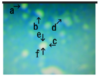
Microscopic appearance of synthetic DNA (Human placenta) crown cells in final sample. Figure 1 shows numerous celllike objects (arrows a, b, c and d). Cell-like objects measured approximately 3-5μm (arrow d). Figure 2 shows a magnification of the structure indicated by arrow a in Figure 1. The colors of the structures facilitate observations and explanations of other figures. However, the different colors also indicate different substances. For convenience, yellow-green structures are called o-structures and orange structures are called m-structures, as described previously [7]. Four types of objects were observed (arrows a, b, c and d). Most objects consisted of o-structures (Figure 2, arrow e) and m-structures (Figure 2, arrow f). Objects (arrows a, b and c) may be not a single cell, judging from their sizes. Objects (arrow b) may also have a narrow structure (arrow h) and may divide to form two cell-like objects.
Figure 1: Microscopic appearance of synthetic DNA (Human placenta) crown cells in final sample. Numerous cell-like objects (arrows a, b, c and d) are shown. Cell-like objects measured approximately 3-5 μm (arrow d).
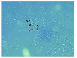
Figure 2:Magnification of the structure shown by arrow a in Figure 1. Four objects are shown (arrows a, b, c and d); Most objects are yellow-green (o-structures; arrow e) or orange in color (m-structures; arrow f); Some objects (arrow b) have a narrow region (arrow h); The object shown by arrow d appeared similar to a typical cell and was connected to other objects (arrows b and k).
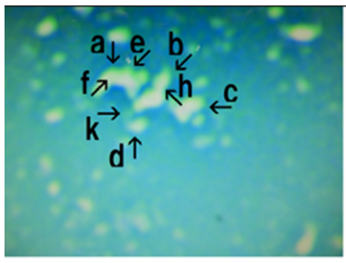
Other objects (arrow d) appeared similar to a typical cell. Cells may be derived from objects (arrow b), because the cells (arrow d) are connected to objects (arrow b, k) that consist of o-structures. Figure 3 shows a magnification of the structure shown by arrow b in Figure 1. Several cell-like objects were observed (arrows a, b, c, d, e and f). These objects consisted of o-structures (Figure 3, arrow h), which enclosed m-structures (arrow k). Objects (arrow a, b, c and d) may be not a single and may give rise to new cells. For example, elongation of an o-structure (arrow i) was observed in one object (arrow c). Further, it is possible that a cell developed from the o-structure indicated by arrow i in Figure 3. New or mature cells are shown in Figure 3 (arrows e and f, respectively). Figure 4 shows a magnification of arrow c in Figure 1.
Figure 3:Magnification of the structure shown by arrow b in Figure 1. Several cell-like objects are shown (arrows a, b, c, d, e and f); These objects consist of o-structures (arrow h), which enclosed m-structures (arrow k).
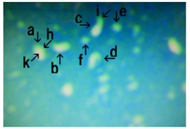
Figure 4:Magnification of the structure shown by arrow c in Figure 1. Several cell-like objects are shown (arrows a, b, c, d, e and f); An object (arrow a) consisting of an o-structure (arrow k) and m-structure (arrow h).
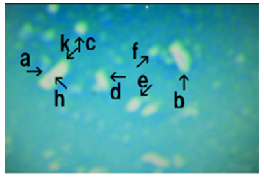
Several cell-like objects were observed (arrows a, b, c, d, e and f). One object (arrow a) consisted of an o-structure (arrow k) and an m-structure (arrow h). Single cell-like objects were observed (arrows c and f). One object (arrow f) may have developed from the object shown by arrow b. Figure 5 shows a magnifications of the structure shown by arrow d in Figure1. Several cell-like objects were observed (arrows a, b, c and d). Also, object (arrow c) consisting of an o-structure (arrow e) and an m-structure (arrow f) was observed. Objects similar to single cells were observed (arrows a, b and c). These objects were covered with o-structures. In previous experiments [4], synthetic DNA (E. coli) crown cells were cultured for seven generations over approximately 50 days, and many cells and related objects were observed in the culture medium. The findings showed that synthetic DNA (E. coli) cells could be cultured for an extended period. On the other hand, it took a long time to obtain such a strain and it may be possible to establish a strain of synthetic DNA crown cells in a more convenient manner, such as by decreasing the culture time.
Figure 5:Magnification of the structure shown by arrow d in Figure 1. Several cell-like objects are shown (arrows a, b, c and d); An object (arrow c) consisting of an o-structure (arrow e) and m-structure (arrow f).
In the present experiments with DNA (Human placenta) crown cells, cells were first cultivated in egg white and DMEM. Many cells and related objects were observed after culturing for about 30 days. The cells obtained in this way may then be cultivated and certified as a strain. In this way, a strain of cells was/could potentially be obtained within 30 days of culture. On the other hand, although studies on the culture medium were not conducted, egg white was used as the medium for the first culture because synthetic DNA crown cells have been shown to multiply in egg white in vivo [2,5,6]. In subsequent cultures, DMEM was used as the cells that were prepared from cultured cells could be cultured in DMEM [8]. It is thus unclear whether the present culture medium is optimal for culturing synthetic DNA crown cells.
On the other hand, it could be convenient to use such a medium as it would permit the development of a strain of synthetic DNA crown cells within 30 days. It was therefore unclear whether it is necessity to carry out the third culture to obtain a strain. Hence, even if more effective and convenient methods could be used to culture synthetic DNA crown cells, then the present method could be used as a standard protocol for preparing strains of synthetic DNA crown cells. On the other hand, it was unclear how the cultured synthetic DNA crown cells multiply. During the cultivation of synthetic DNA crown cells, various kinds of objects were observed (Figure 2, arrows a, b and c). These objects may be not cells, but objects that develop into new cells. Most of the cultured objects consist of o-structures (yellow-green color) and m-structures (orange color). Since the outside of DNA crown cells consists of SPH-DNA or related substances, these o-structures may correspond to such components. In the case of cell multiplication, such components may grow or elongate [4].
However, it is currently not clear how o-structures (SPH-DNArelated components) grow or elongate. Recently, it was demonstrated that some objects, possibly SPH-DNA-related components of synthetic DNA crown cells, became elongated in the presence of adenosine-DNA [9]. The findings showed that SPH-DNA associated components were elongated without any enzymes, suggesting that synthetic DNA crown cells were successfully cultivated. The finding implies that non-living structures can develop into organisms. On the other hand, many objects consisting of o-structures and m-structures that may develop into cells were observed during culture (Figure 3, arrows a, b and c). Many challenges remain to prove that synthetic DNA crown cells can develop into a strain, i.e., change from inorganic substances to organic substances. For example, what are the objects that are source of new cells (Figure 3, arrows a, b, and c) and how can they be observed after 30 days of culture? These points need to be clarified. Moreover, methods for producing a growth curve for cultured synthetic DNA crown cells have not yet been established. Cell growth was not confirmed by microscopy, because it was unclear whether single cultured cells would be round or irregular, or whether they have a cell cycle. In a previous study on estimating the growth of DNA crown cells [10], the amount of DNA in culture medium was measured and it was demonstrated that cells developed. Such methods can be used to estimate the growth of cultured synthetic DNA crown cells.
This study described a protocol for prepare a strain of synthetic DNA crown cells. To better clarify the difficulties associated with culturing synthetic DNA crown cells, more synthetic DNA crown cell strains need to be developed in future [11].
Conclusion
The present study described a protocol for prepare a strain of
synthetic DNA crown cells using synthetic DNA (Human placenta)
crown cells. The procedures are as follows
a) A total of 20μL of sample (synthetic DNA crown cells) was
added to 200μL of egg white and the mixture was incubated
for 7 days at 37 °C (primary culture).
b) Then, 50μL of sample was added to 500μL of DMEM and the
mixture was incubated for 7 days at 37 °C (secondary culture).
c) Further, 50μL of sample was added to 500μL of DMEM and the
mixture was incubated for 7 days at 37 °C (third culture).
To scale up, 0.5mL of sample was added to 5.0mL of DMEM and the mixture was incubated for 7 days at 37 °C (fourth culture).
References
- Inooka S (2012) Preparation and cultivation of artificial cells. App Cell Biol 25: 13-18.
- Inooka S (2016) Preparation of artificial cells using eggs with sphingosine-DNA. J Chem Eng Process Technol l7: 277.
- Inooka S (2016) Aggregation of sphingosine-DNA and cell construction using components from egg white. Integrative Molecular Medicine 3(6): 1-5.
- Inooka S (2022) Preparation of a DNA ( coli) crown cell line in vitro-microscopic appearance of cells. Annals of Reviews and Research 28(1).
- Inooka S (2017) Biotechnical and systematic preparation of artificial cells. The Global Journal of Researches in Engineering 17(1-C).
- Inooka S (2017) Systematic preparation of artificial cells (DNA crown cells). J Chem Eng Process Technol 8: 327.
- Inooka S (2022) Microscopic appearance of synthetic DNA ( coli) crown cells in primary culture. App Cell Biol Japan 35: 71-98.
- Inooka S (2000) Cyto-organisms (cell-originated cultivable particles) with sphingosine-DNA Comm. Appl Cell Biol 17: 11-34.
- Inooka S (2023) Formation and microscopic appearance of fireworks-like objects created from synthetic DNA ( coli) crown cells with adenosine-DNA (E. coli). Annals of Review and Research 8 (2): 555760.
- Inooka S (2013) Preparation of artificial cells for the yogurt production. App Cell Biol 26: 13-17.
- Inooka S (2022) Microscopic appearance of synthetic DNA ( coli) crown cells in secondary cultures. Novel Research in Science 12(3).
© 2023 Shoshi Inooka. This is an open access article distributed under the terms of the Creative Commons Attribution License , which permits unrestricted use, distribution, and build upon your work non-commercially.
 a Creative Commons Attribution 4.0 International License. Based on a work at www.crimsonpublishers.com.
Best viewed in


.jpg)


 Editorial Board Registrations
 Submit your Article
 Refer a Friend
 Advertise With Us
.jpg)


.jpg)


.bmp)
.jpg)
.png)
.jpg)


.jpg)


.png)

.png)


.png)


