- Submissions

Full Text
Integrative Journal of Conference Proceedings
Muculent and Soggy-Myxoid Liposarcoma
Anubha Bajaj*
A.B. Diagnostics, Panjab University, India
*Corresponding author:Anubha Bajaj, A.B. Diagnostics, Panjab University, A-1, Ring Road, Rajouri Garden, New Delhi -110027, India
Submission: June 14, 2023;Published: June 21, 2023

Volume3 Issue4June , 2023
Opinion
Myxoid liposarcoma appears as malignant tumour constituted of primitive, non-lipogenic mesenchymal cells and signet ring lipoblasts enmeshed within a prominent myxoid stroma characteristically incorporated with branching vascular configurations. Myxoid liposarcoma exhibits a repetitive molecular alteration with chromosomal translocation t(12;16)(q13;p11.2) with FUS-DDIT3 genetic fusion or translocation t(12;22)(q13;q12) with EWSR1-DDIT3 genomic rearrangement. Majority of tumour parenchyma is non lipogenic and is pervaded with scattered lipoblasts with characteristic signet ring histology. Surrounding prominent myxoid stroma with branching vasculature is designated as ‘chicken wire’ vasculature. High grade lesions, previously designated as ‘round cell’ liposarcoma demonstrate a propensity for initial distant metastasis into soft tissue or bony site, as documented with singular extremity to contralateral extremity, retroperitoneum or spine. Myxoid liposarcoma is additionally designated as round cell liposarcoma as the neoplasms depict identical molecular alterations with various transitional morphological configurations. Myxoid liposarcoma is designated as high-grade tumour or low grade tumour contingent to proportionate round cell component. As per World Health Organization (WHO) classification, high grade lesions manifest >>5% round cell component. Borderline neoplasms with round cell component <5% are designated as lesions with foci of ‘transition’ which demonstrate an ambiguous diagnostic significance.
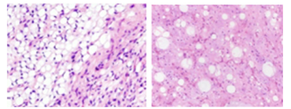
Myxoid liposarcoma represents ~5% of adult sarcoma. Disease incidence is predominant within fourth decade to fifth decade although pediatric population may display the condition. Elderly subjects are infrequently incriminated. A specific gender predilection is absent. Commonly, tumefaction is encountered upon extremities of incriminated young adults. Proximal thigh is commonly implicated. Primary retroperitoneal neoplasms are infrequently observed. However, metastasis into retroperitoneal soft tissue is commonly exemplified. Myxoid liposarcoma represents a propensity for multifocal disease. Hematogenous tumour dissemination may ensue although pulmonary parenchyma is spared. Besides, primary subcutaneous tumours are documented. Majority of myxoid liposarcomas exemplify chromosomal translocation t(12;16)(q13;p11.2) with FUS-DDIT3 genetic fusion. Exceptionally, chromosomal translocation t(12;22)(q13;q12) with EWSR1-DDIT3 genetic fusion may be discerned. Generally, genetic rearrangements involve several chromosomes and appear complex with and involve other chromosomes. Upon cytological examination, myxoid substances appear inundated with arborizing vascular articulations and lipoblasts. Grossly, the neoplasm characteristically appears multi-nodular and well circumscribed. The cut surface of low-grade neoplasms appears gelatinous whereas high grade tumours appear solid and fleshy. Upon microscopy, low grade myxoid liposarcoma appears as a pauci-cellular neoplasm composed of uniform, monomorphic, stellate or fusiform cells. Cellular and nuclear atypia or significant pleomorphism is absent. A prominent plexiform vascular network constituted of delicate, thin walled, arborizing, curvilinear capillaries akin to ‘chicken wire’ fencing enmeshes the tumour cell clusters. Innumerable signet ring lipoblasts appear to congregate upon periphery of tumour lobules, thereby configuring a lipoblastoma-like appearance. Encompassing mucoid matrix is preponderantly composed of hyaluronic acid which configures enlarged mucoid pools, simulating ‘pulmonary oedema pattern. Mucoid matrix can be highlighted with stromal mucin stains as Alcan blue. Foci of metaplastic cartilage or bone can be exceptionally discerned. Metaplastic tissue retains the pre-existing molecular alterations in the absence of progressive molecular modifications. However, the aforesaid foci may not represent dedifferentiation of neoplastic tissue. Mitotic activity is minimal. Low grade myxoid liposarcoma manifests with several, exceptionally discerned morphological variants. High grade myxoid liposarcoma exemplifies compact aggregates of round cells. The hyper-cellular neoplasm is composed of solid sheets or compact aggregates of primitive or round cells permeated with minimal eosinophilic cytoplasm, a component which exceeds >5% of tumour tissue sample. Enlarged foci of mature adipocytic differentiation may be encountered (Figure 1). TNM staging of soft tissue sarcoma confined to trunk and extremities as per American Joint Committee on Cancer (AJCC) 8th edition [1,2].
Figure 1:Myxoid liposarcoma.

Primary tumour
TX: Tumour cannot be assessed, T0: No evidence of primary tumour, T1: Tumour <5 centimeters in greatest dimension, T2: Tumour ≥5 centimeters and <10 centimeters in greatest dimension, T3: Tumour ≥10 centimeters and <15 centimeters in greatest dimension, T4:Tumour >15 centimeters in greatest dimension.
Regional lymph nodes
NX: Regional lymph nodes cannot be assessed, N0: Regional lymph node metastasis absent, N1: Regional lymph node metastasis present.
Distant metastasis
M0: Distant metastasis absent, M1: Distant metastasis present into sites such as pulmonary parenchyma French Federation of Cancer Centers Sarcoma Group (FNCLCC) classifies adult sarcoma as tumour differentiation ~score 1: sarcoma simulates adult normal tissue ~score 2: sarcoma with confirmed histological diagnosis ~score 3: Poorly differentiated sarcomas as embryonal sarcoma, synovial sarcoma, epithelioid sarcoma, clear cell sarcoma, soft tissue alveolar sarcoma, undifferentiated sarcomas or sarcomas of uncertain histological subtype mitotic index ~score 1: 0-9 mitoses per 10 high power fields ~score 2: 10-19 mitoses per 10 high power fields ~score 3: >19 mitoses per 10 high power fields tumour cell necrosis ~score 1: absence of tumour necrosis ~score 2: <50% of tumour appears necrotic ~score 3: >50% of tumour appears necrotic. Histologic grade of soft tissue sarcoma of trunk and extremities [1,2].
GX: Tumour grade cannot be assessed G1: Tumour differentiation, mitotic count and necrosis quantified between total score of 2 and 3 and minimal metastasis G2: Tumour differentiation, mitotic count and necrosis quantified between total score of 4 and 5 with possible metastasis G3: Tumour differentiation, mitotic count and necrosis quantified between total score of 6,7 or 8 with significant possibility of metastasis. Tumour stages of soft tissue sarcoma of trunk or extremities(3,4), stage IA: T1, N0, M0, GX or G1, stage IB: T2, T3, T4, N0, M0, GX or G1, stage II: T1, N0, M0, G2 or G3, stage IIIA: T2,N0, M0, G2 or G3, stage IIIB:T3, T4, N0, M0, G2 or G3, stage IV: Any T, N1, M0, any G OR any T any N, M1 any G. Myxoid liposarcoma appears immune reactive to vimentin or S100 protein. Myxoid liposarcoma appears immune non-reactive to CD34 or MDM2. Low grade myxoid liposarcoma requires segregation from neoplasms such as atypical lipomatous tumour/well differentiated liposarcoma (ALT/WDL), Ext skeletal myxoid chondrosarcoma, lipoblastoma/lipoblastomata’s, myxoid dermatofibrosarcoma protuberans or myxoma. High grade myxoid liposarcoma requires demarcation from neoplasms such as myxofibrosarcoma or pleomorphic liposarcoma. Additionally, myxoid liposarcoma necessitates segregation from round cell sarcomas as Ewing sarcoma, BCOR- CCNB3 rearranged sarcoma or CIC-DUX4 rearranged sarcoma.
Although several neoplasms exhibit plexiform vasculature and aggregates of signet ring cells, myxoid liposarcoma indicated upon clinical or radiological grounds may be confirmed with molecular evaluation.
Miniature tissue samples can be subjected to molecular assessment as fluorescent in situ hybridization (FISH), adopted for detecting FUS or EWSR1 genetic rearrangement. The rearrangements may be appropriately discerned by fluorescent in situ hybridization (FISH), polymerase chain reaction(PCR), classic cytogenetics or next generation sequencing. Myxoid liposarcoma with a predominant round cell component exhibits a propensity for distant metastasis. However, distant metastases may ensue following decades of tumour emergence, thereby necessitating extended tumour monitoring. Pure, low grade myxoid liposarcoma appear minimally aggressive and demonstrate a penchant for tumour reoccurrence wherein distant metastasis ensues in up to 10% instances. Myxoid liposarcoma enunciating the exceptional TP53 or CDKN2A genetic mutation is accompanied by inferior prognostic outcomes. Magnetic resonance imaging (MRI) can optimally detect high grade neoplasms. Besides, antecedent discernment of distant metastasis may be achieved with cogent imaging studies [3-5].
References
- Suardi D, Hamdani Z, Palungkun IG, Jessica K, Kemala IM, et al. (2022) Myxoid liposarcoma of the Vulva: A rare malignancy mimicking benign vulvar mass. Am J Case Rep 23: e937575.
- Mokfi R, Boutaggount F, Maskrout M, Ghizlane R (2022) Giant mesenteric myxoid liposarcoma: Challenges of diagnosis and treatment. Radiol Case Rep 17(11): 4227-4231.
- Yu JI (2022) Myxoid liposarcoma: A well-defined clinical target variant in radiotherapy for soft tissue sarcoma. Radiat Oncol J 40(4): 213-215.
- Sassi F, Sahraoui G, Charfi L, Zemni I, Karima M, et al. (2022) A rare case report of a myxoid liposarcoma arising from the broad ligament. Rare Tumors 14: 20363613221148839.
- Tuzzato G, Laranga R, Ostetto F, Elisa B, Giulio V, et al. (2022) Primary high-grade myxoid liposarcoma of the extremities: Prognostic factors and metastatic pattern. Cancers (Basel) 14(11): 2657.
© 2023 Anubha Bajaj. This is an open access article distributed under the terms of the Creative Commons Attribution License , which permits unrestricted use, distribution, and build upon your work non-commercially.
 a Creative Commons Attribution 4.0 International License. Based on a work at www.crimsonpublishers.com.
Best viewed in


.jpg)


 Editorial Board Registrations
 Submit your Article
 Refer a Friend
 Advertise With Us
.jpg)


.jpg)


.bmp)
.jpg)
.png)
.jpg)


.jpg)


.png)

.png)


.png)


