- Submissions

Full Text
Integrative Journal of Conference Proceedings
Experience in Using Synchrotron Radiation for Structural Researches of Biological Systems
Korneev VN1* and Lanina NF2
1Institute of Cell Biophysics, RAS, Pushchino, Russia
2Institute of Theoretical and Experimental Biophysics, RAS, Pushchino, Russia
*Corresponding author: Korneev VN, Institute of Cell Biophysics, RAS, Pushchino, Russia
Submission: April 01, 2021;Published: April 22, 2021

Volume2 Issue3April, 2021
Introduction
Studying the structural mechanism of the functional activity of biological systems, an
important role belongs to the instrumental and methodical developments intended for the
energy-dispersive (Θ=const) and monochromatic (λ=const) diffractometry using Synchrotron
Radiation (SR). To solve the problems of structural biology of tissues, the SR of two Russian
centers of collective use-the Siberian and Moscow regions-was used. X-ray stations were
created on beam-lines of storage ring VEPP-3 (SCSTR, Novosibirsk) and Siberia-2 (NRC
«Kurchatov Institute», Moscow). Experimental techniques and research methods were
intended for the following experiments: the study of the structure and structural dynamics
of cross-striated skeletal muscle; the structural study of epithelial tissue samples in normal
and pathological conditions; the study of natural and engineering silk constructions, as well
as the effect of high-frequency electrosurgical welding (HF-welding) on biological tissues.
Such heterogeneous multicomponent ensembles of living systems are characterized by a high
order of structure-periodicity in the nanometer range of about 1-100nm, and weak scattering
ability due to their extremely low concentration in native tissues [1].
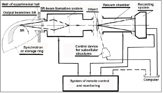
X-ray stations based on the methods of monochromatic (λ=const) (FRAKS, DICSI)
and energy (Θ=const) (KEMUS) diffractometry were created on the working channels of
SR sources. Figure 1 shows their generalized block diagram with fundamental differences
for each method in the systems of SR beam formation and registration of X-ray diffraction
patterns based on position-sensitive coordinate or energy-dispersion detectors.
Figure 1:

New approaches to the formation of the SR beam are implemented at the stations. Thus, for the λ=const method, the following operations were performed: a reversed version of the arrangement of X-ray optical zoom lenses based on a crystal-monochromator and polysection mirrors; high-speed X-ray diffractometry with simultaneous registration of micron periodicity of the structure in the visible range of the spectrum; remote-controlled methods of combining the SR beam with the object and adjusting the station with minimal background illumination. For the Θ= const method, some modifications were performed: registration in both the meridional (interval 0-50mrad) and sagittal (interval 0-25mrad) directions with an accuracy of 0.05mrad; collimating device with a mirror zoom; object cooling and scanning. These modifications allowed to increase the signal-to-noise ratio by 6-7 times.
This equipment was created on the basis of the block-modular principle, i.e. when the main devices of the stations could be used as independent modules in any experimental scheme. Despite the different nature of the tasks to be solved, much of the experimental equipment was unified, for example, such units as monochromatic and mirror zoom lenses, primary beam collimators, as well as devices for vacuuming, remote control of optical system components and software.
In experimental installations, an original sequence of crystalmirror focusing X-ray optical elements located in vacuum-operated modules was created, which made it possible to increase the light intensity of the beam-line, reduce radiation and thermal loads on the mirror surfaces of the elements, and also provide suppression of the higher harmonics of the main wavelength. For monochromatization and focusing of the SR beam in the horizontal plane, the traditional scheme of reflection of the X-ray beam from a curved crystal according to the Johann geometry is applied.
Monochromatization of the polychromatic beam coming out of the storage ring is carried out by a device with a crystalmonochromator, which also serves as a transfocator for focusing the beam in the sagittal (horizontal) plane. In such a monochromatic transfocator, crystals with a high coefficient of suppression of even harmonics (germanium, silicon) were used, which makes it possible to exclude high-energy components from the diffracted beam with the help of a subsequent mirror transfocator, which is based on a polysectional system of mirrors of total external reflection and focuses the beam in the meridional plane.
This section presents experimental results that characterize the possibilities of instrumental and methodological support for X-ray diffraction studies of biological objects and other samples using various recording systems on SR channels.
Figure 2 shows the changes in the diffraction pattern during the functioning of the biological system by the example of the study of alive frog sartorius muscle using the “diffraction cinema” technique with the OD-3 detector at a sample-detector distance of 1500mm and λ= 0.16nm. The figure shows the kinetics of the intensity of equatorial reflections in the dynamic process of changing the muscle structure. A set of X-ray patterns from one cycle of isometric contraction (12 frames with a duration of 10ms each) is given. In the third frame, the muscle was stimulated by a 3ms pulse. Then the intensities of reflections (11) and (10) are recorded. There is an increase and decrease in the intensity, respectively. On the twelfth frame, you can see that the muscle is returning to its original state. Thus, the stimulus can be considered as a tool for a directed change in the nature of the contraction in different phases of the active state of the muscle.
Figure 2:

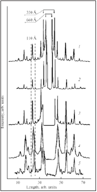
Figure 3 shows comparative radiographs obtained from collagen fibers from the rat tail tendon, which served as a test sample. The registration was carried out on the same coordinate by different detectors at a wavelength of λ= 0.15nm. Using the OD-3 (1) detector under experimental conditions: The storage current I = 52mA, the exposure T = 60s, the sample–detector distance L = 1500mm, a higher resolution of the diffraction lines is obtained and more than 14 orders of reflection are clearly visible in the range from about 66 nm. Similar results are viewed on the linear profile of the diffraction pattern obtained on Image Plate (3) at I = 125mA, T = 20s, L = 500mm. X-ray patterns obtained using systems based on cooled matrices show a minimal background with a good spatial resolution when using the SX-165 (2) detector (I = 71mA, T = 20s, L = 2440mm) compared to systems with ICX 249 (4) and ICX 285 (5) matrices and gadolinium screens (Gd2O2S:Tb) at L = 500mm and I = 83mA, T=300s and I= 84mA, T = 600s, respectively.
Figure 3:

Figure 4 shows a small-angle diffraction pattern of collagen. A part of the image is shown in the meridional direction (a) (for clarity, the picture is located horizontally), and the profile of the reflections along the axis (b) is shown. On the X-ray image obtained at the DICSI station using the SX-165 detector (λ = 0.165nm, L = 2440mm, I = 71mA, T = 20s), 11 orders of reflection from the main period of the structure of 66 nm are observed in the range of registration angles 2θ ≈ 0.14°-1.57°. The presented results of the developed hardware and methodological support made it possible to use SR to study the vital activity of an organism at different levels of the biological organization of structures.
Figure 4:

References
- Korneev VN, Shlektarev VA, Zabelin AV, Lanina NF, Tolochko BP, et al. (2019) Instrumental and methodological developments for the study of biological structures using synchrotron radiation. Russian Journal of Biological physics and Chemistry 4(2): 259-269.
© 2021 Korneev VN. This is an open access article distributed under the terms of the Creative Commons Attribution License , which permits unrestricted use, distribution, and build upon your work non-commercially.
 a Creative Commons Attribution 4.0 International License. Based on a work at www.crimsonpublishers.com.
Best viewed in


.jpg)


 Editorial Board Registrations
 Submit your Article
 Refer a Friend
 Advertise With Us
.jpg)


.jpg)


.bmp)
.jpg)
.png)
.jpg)


.jpg)


.png)

.png)


.png)


