- Submissions

Full Text
Gerontology & Geriatrics studies
Radial Artery pseudo aneurysm with Thrombosis- A Case without Trauma or Anterior Local Surgery
Márcio Luís Duarte* and Élcio Roberto Duarte*
Hospital Irmã Dulce, Praia Grande, São Paulo, Brazil
*Corresponding author: Márcio Luís Duarte, Hospital Irmã Dulce, Praia Grande, São Paulo, Brazil
Submission: December 23, 2017; Published: January 10, 2018

ISSN : 2578-0093Volume1 Issue4
Abstract
A 69 years- old man with right wrist pain and functional limitations for 06 months. Finkelstein, Tineland Phalentests were negative. Import antedema in the wrist and hand at physical examination. Ultrasonography demonstrated pseudo aneurysm with thrombosis in the distal radial artery.
Keywords: Aneurysm, False/diagnosticimaging; Ultrasonography, Doppler; Radial artery
Introduction
pseudo aneurysm of the upper extremity is rarer as compared to the lower extremity [1]. Pseudoaneurysms of the radial artery are uncommon [2] and they are morphologically characterized as fusi formors acculardilatations, probably representing a form of dissection, with rupture of the artery between media and adventitial layers of the vascular wallor resulting from weaknes soft headventitia itself with subsequenten capsulation of the perivascular hematoma. Sometimes it is contained in adventitia or adjacent connectivet issues, promoting local flow, and can cause a series of complications [3].
An arterial pseudoaneurysmor false aneurysms are shaped commonly by penetrating trauma of a native vessel, proceed by hemorrhage and extravasation. Although the detection of such complications may result within hours from the time of insult, they may occur one to several months afterward [2].
Case Report
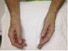
Figure 1: Physical examination comparing wrists and hands, demonstrating pronounced right edema

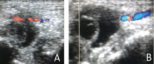
A 69 years old man with right wrist pain and functional limitations for 06 months. Finkelstein, Tineland Phalentests were negative. Important edema in the wrist and hand at physical examination (Figure 1). Ultrasonography demonstrated pseudo aneurysm with thrombosis in the distal radial artery (Figures 2 & 3).
Figure 2: Ultrasonography without Doppler demonstrating thrombosis in the distal radial artery (white arrow).

Figure 3: Ultrasonography with Doppler in A and B demonstrating pseudo aneurysm with thrombosis in the distal radial artery.

Discussion
pseudo aneurysm following arterial injuries are rare occurrences, but they are described in the literature after vascular access attempts to arteriovenous fistulae, catheterization of arteries, arterial blood gas analysis, and other invasive procedures [2]. Normally pseudoaneurysmis associated with multiple attempts of cannulation. May be using an ultrasound for arterial cannulation in high-risk patients might reduce the incidence of pseudoaneurysmsas the number of attempts for cannulation is reduced significantly [4].
Depending on the size of the aneurysmsac, a mass might not be palpated leaving pain as the lonely symptom. If a mass can be palpated, it may be pulsatile, but it may not have a thrill. At the physical examination of a pseudo aneurysm, Allen's test is generally negative- arterial pulses are usually palpated distal to the mass. Morbidity can be severe and is related to distal embolization from microemboli, venous compression orevenfrank rupture [2].
There are three characteristic features of pseudoaneurysms on ultrasound [2]
The presence of expansilepulsatility, detection of turbulent flow that appears as a classic "yin-yang" sign, and a hematoma with variable echogenicity.
A. The variable echogeni city can represent separate episodes of bleeding and rebleeding.
B. The identification of a "to-and-fro" spectral wave form within the neck is considered pathognomonic for a pseudo aneurysm.
Ultrasound compression therapy involves direct ultrasound- guided squeeze of a pseudo aneurysm to obstruct the inflow tract of blood. The stasis of blood promotes coagulation causing occlusion and resolution of the pseudo aneurysm. Unluckily, compression must behold for over an hour to obtain occlusion- sometimes longer for anticoagulated patients [2].
Surgical intervention may need to be considered if the complete occlusion is not achieved. Another option for occlusion of pseudoaneurysms involves ultrasound-guided thrombin injection in to the sac [2].
When progressive thrombosis of the pseudo aneurysm can be demonstrated and there is no enlargement of the pseudo aneurysm and no clinical evidence of pain or neuropathy, such findings port end that the pseudo aneurysm has a tendencyto self-heal [5].
Conclusion
Pain can be caused by vascular diseases and it's something that the sonographer doesn't research unless it's on suspicion of requesting physician. The sonographer should look for vascular diseases when there's pronouncededema and pain.
References
- Kakar BK, Babar KM, Khan A, Naeem M, Khan I, et al. (2006) Surgical experience of post-traumatic pseudo aneurysm of the brachial artery at a tertiary care hospital. JPMA 56(9): 409-412.
- Pero T, Herrick J (2009) pseudo aneurysm of the radial artery diagnosed by bedside ultrasound. West J Emerg Med 10(2): 89-91.
- Romanus AB, Mazer S, Neto AC, Liu CB, Toni FS, et al. (2002) Pseudo- aneurismas- relato de dois casos e revisao da literatura. Radiol Bras 35(5): 303-306.
- Ranganath A, Hanumanthaiah D (2011) Radial artery pseudo aneurysm after percutaneous cannulation using Seldinger technique. Indian J Anaesth 55(3): 274-276.
- Kotval OS, Khoury A, Shah PM, Babu SC (1990) Doppler sonographic demonstration of the progressives pontaneous thrombosis of pseudoaneurysms. J Ultrasound Med 9(4): 185-190.
© 2018 Márcio Luís Duarte, et al. This is an open access article distributed under the terms of the Creative Commons Attribution License , which permits unrestricted use, distribution, and build upon your work non-commercially.
 a Creative Commons Attribution 4.0 International License. Based on a work at www.crimsonpublishers.com.
Best viewed in


.jpg)


 Editorial Board Registrations
 Submit your Article
 Refer a Friend
 Advertise With Us
.jpg)


.jpg)


.bmp)
.jpg)
.png)
.jpg)


.jpg)


.png)

.png)


.png)


