- Submissions

Full Text
Experimental Techniques in Urology & Nephrology
Sarcomatoid Carcinoma of Kidney: Case Report
Yddoussalah O*, Touzani A, Karmouni T, Elkhader K, Koutani A and IBN Attya Andaloussi A
Department of Urology B, Mohamed V University, Morocco
*Corresponding author: Othmane Yddoussalah, Department of Urology B, CHU Ibn Sina, Faculty of Medicine and Pharmacy, Mohamed V University, Rabat, Morocco
Submission: December 24, 2017; Published: January 18, 2018
ISSN: 2578-0395 Volume1 Issue3
Abstract
Sarcomatoid renal carcinoma is a rare and agressive variant of the cancer of the kidney. This tumor are indifferencied and originates from all renal cell carcinomas. The diagnosis is exclusively histologic and therapeutic modalities are limited to radical nephrectomy. We report a new case and will discuss diagnosis, therapeutic and prognostic characteristics of this rare and agressive entity.
Keywords: Sarcomatoid carcinoma; Kidney; Prognosis; Surgery
Introduction
The term sarcomatoid renal cell carcinoma was introduced by Farrow in 1968, who studied 38 cases of kidney tumors with a double contingent of malignant cells: a carcinomatous component and a pleomorphic undifferentiated component of sarcomatoid-like cells [1]. The classification of tumors with features of "carcinoma" and "sarcoma" has been debated since Virchow in 1864, which initiated the term carcinosarcoma [2]. It is currently accepted that sarcomatoid carcinomas of the kidney are not a distinct histological entity, but may develop from all histological subtypes of renal cell carcinomas, as proposed in the WHO 2004 classification [3,4]. Sarcomatoid transformation of renal cell carcinoma seems uncommon (1 to 15%), which explains the low number of series in the literature.
From its description, the pejorative prognosis ofthe sarcomatoid contingent has been clearly established with respect to conventional renal cell carcinoma [2]. The aim of this article was to carry out a review based on the recent literature of epidemiological, clinical- biological, prognostic and therapeutic sarcomatoid carcinomas of the kidney.
Observation
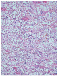
Mr. K.S, 70 years old, with no pathological history. She complained for four months of low back pain associated with a single episode of hematuria, without other urinary disorders, or digestive associated. On clinical examination, the patient was afebrile. A mass was palpated at the left flank extending 4cm below the costal margin, solid consistency. Computed tomography showed a higher mass of the 14cm long axis, which had a tissue density, which increased in an inhomogeneous manner after injection of the contrast medium. This mass shows a renal pedicle break-in and invasion of the renal pedicle (Figure 1). In view of the suspicion of invasion of the renal pedicle, a magnetic resonance imaging supplement was performed to show an aggressive renal process with thrombotic material occupying the left renal vein with the IVC is free (Figure 2). The surgical approach was left sub costal. After detachment of the left colic angle at the exploration we discovered a voluminous tumor adherent to the adjacent structure and in particular the adrenal We had proceeded to a left enlarged nephrectomy, before the invasion of the left renal vein an extraction of the thrombus tumor by veinotomy was realized. On gross examination, the nephrectomy piece, measuring 18 x 13 x 8cm and weighing 1kg, found a 13cm beige-white tumor infiltrating perirenal fat. It is the site of extensive tumor necrosis estimated at 60% of the tumor area. The vein lumen is filled by a tumor thrombus. Microscopic examination revealed a malignant tumor proliferation made of fusiform cells, arranged in intersecting bundles associated with numerous figures of mitosis. Presence of pure-tumor vascular emboli (Figure 3). This morphological analysis concluded Furhman's nuclear grade 4 sarcomatoide carcinoma. Class pT3aNx.
Figure 1: Tomodensitometric section showing superior polar mass of the left kidney with injection of the heterodense contrast medium. This process measures 140x83mm of large and necrotic zone seat.
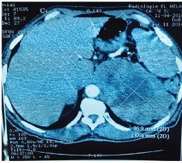
Figure 2: Sagittal magnetic resonance imaging showing a left renal process, infiltrating the renal sinus and the renal pedicle. The VCI remains free and of normal caliber.
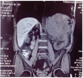
Figure 3: Histological aspect (HES, x 400): mixed appearance with the tumor is made of fusiform cells sometimes irregular nucleus.
Discussion
Sarcomatoid carcinoma of the kidney is a rare variant of kidney cancer. Its incidence in contemporary studies is estimated to be between 1 and 13% of all renal tumors [5]. The median age of these patients, between 55 and 60 years of age (32-87 years), was comparable to renal cell carcinomas without sarcomatoid transformation [6,7].
The sex ratio did not seem not different with about twice as many men as women. Sarcomatoid kidney carcinomas were frequently found on clinical signs (85 to 90% of cases after the series), whereas renal carcinomas without sarcomatoid differentiation with more than 50% chance of discovery [2,8-10]. The most common symptoms were lower back pain or hematuria. Microscopically It is about bulky masses, with numerous haemorrhagic and necrotic foci. This tumor often extends into the perirenal fat and invades the vessels of the hilum. Sarcomatoid carcinoma is a very aggressive, discovery often at an advanced stage of the TNM classification of kidney cancer. Below the detailed TNM classification for kidney cancer variant (Table 1).
Table 1: TNM staging of renal cell carcinoma.
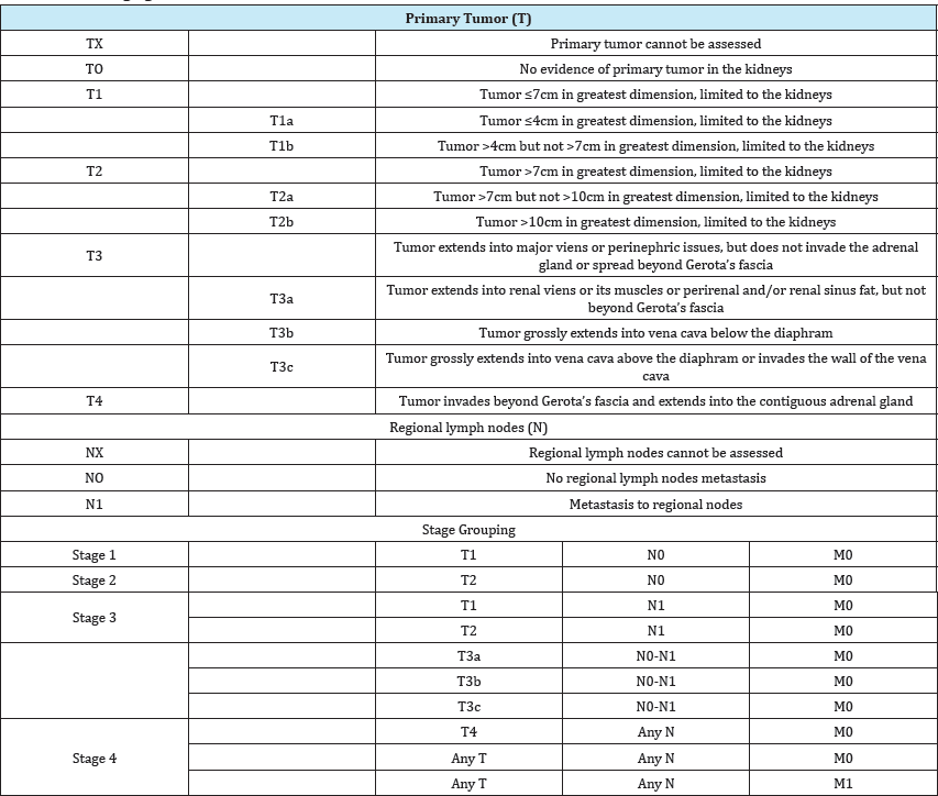
Sarcomatoid carcinoma of the kidney is frequently found in a metastatic stage. The usual sites of sarcomatoid carcinoma metastases are the lung, bone, liver, ganglia, and brain [8,11]. Microscopically, it is a mixed-component tumor, comprising varying degrees of pseudo-sarcomatous elements and malignant epithelial elements [12]. The sarcomatoid beaches are sometimes so dense that it is difficult to assert the epithelial nature of the lesion. It is therefore necessary to multiply the cutting plans. Immunohistochemicals ca be of great help in these cases since tumor cells express cytokeratin in 94% of cases, EMA in 50% of cases and vimentin in 56% of cases [6]. Cytologically, they are classified into Furhman 4 nuclear grade. We find below the complete Fuhrman grading of kidney cancer (Table 2).
Table 2: Complete Fuhrman grading of kidney cancer.

Numerous composite morphologic and nuclear grading systems have been proposed for RCC and although that of the Fuhrman classification have achieved widespread usage, the validity of the grading criteria of this classification has been questioned. In addition, there are few studies that have attempted to validate the Fuhrman system for RCCs beyond that of the clear cell subtype. Recent studies have indicated that grading of papillary RCC should be based on nucleolar prominence alone and that the components of the Fuhrman grading classification do not provide prognostic information for chromophobe RCC.
The overall prognosis of patients with sarcomatoid carcinoma of the kidney is pejorative. In the recent literature, median overall survival ranged from 4.9 to 19 months. The survival at 2 years ranged from 15 to 30% and 5-year survival from 2 to 20% [6,7].
The basis of treatment is nephrectomy (partial or enlarged) when it is feasible [13,14]. To date, there is no standard systemic therapy for patients with sarcomatoid carcinoma of the metastatic kidney. In his retrospective series of 43 patients with sarcomatoid carcinoma of metastatic kidney, Golshayan et al reported a rate of 19% of objective responses to antiangiogenic drugs [8]. Efficacy of anti-antigenic agents appeared to be better in patients with a clear cell carcinomatous contingent and a small percentage of sarcomatoid challenge. The study by Haas et al. [15] in a series of patients, with treatment with doxorubicin associated with gemcitabine, showed two complete responses (survival of Six years and eight years) and a partial answer [15]. Studies of Phase II evaluates the combination of anti-angiogenesis and chemotherapy are ongoing (gemcitabine-sunitinib), (gemcitabine-capecitabine- Bevacizumab)
Conclusion
Sarcomatoid carcinoma of the kidney is not a distinct histological entity, but is derived from all renal cell carcinomas. The natural evolution of these kidney tumors is formidable with a poor spontaneous prognosis of this tumor. Currently In the absence of a recommendation, the management of sarcomatoid carcinoma of the kidney should be that of renal cell carcinoma at high risk.
Consent of Patients
Written informed consent was obtained from the patient s next of kin for publication of this case report and any accompanying images.
References
- Farrow GM, Harrison Jr EG, Utz DC, ReMine WH (1968) Sarcomas and sarcomatoid and mixed malignant tumors of the kidney in adults. I Cancer 22(3): 545-550.
- Chao D, Zisman A, Freedland SJ, Pantuck AJ, Said JW, et al. (2001) Sarcomatoid renal cell carcinoma: basic biology, clinical behavior and response to therapy. In Urologic Oncology: Seminars and Original Investigations. Uro Onco 6(6): 231-238.
- Compérat E, Camparo P, Vieillefond A (2006) WHO classification 2004: tumors of the kidneys. Journal de radiologie 87(9): 1015-1024.
- Lopez-BA, Scarpelli M, Montironi R, Kirkali Z (2006) 2004 WHO classification of the renal tumors of the adults. European urology 49(5): 798-805.
- Mian BM, Bhadkamkar N, Slaton JW, Pisters PW, Daliani D, et al. (2002) Prognostic factors and survival of patients with sarcomatoid renal cell carcinoma. The Journal of urology 167(1): 65-70.
- de Peralta-V, Moch M, Amin H, Tamboli M, Hailemariam P, et al. (2001) Sarcomatoid differentiation in renal cell carcinoma: a study of 101 cases. The American journal of surgical pathology 25(3): 275-284.
- Pal SK, Jones JO, Carmichael C, Saikia J, Hsu J, et al. (2013) Clinical outcome in patients receiving systemic therapy for metastatic sarcomatoid renal cell carcinoma: A retrospective analysis. In Urologic Oncology: Seminars and Original Investigations 31(8): 1826-1831.
- Golshayan AR, George S, Heng DY, Elson P, Wood LS, et al. (2009) Metastatic sarcomatoid renal cell carcinoma treated with vascular endothelial growth factor-targeted therapy. Journal of Clinical Oncology 27(2): 235-241.
- Shush B, Breathlessly G, Shih J, Courante S, Finley D, et al. (2012) Impact of pathological tumour characteristics in patients with sarcomatoide renal cell carcinoma. BJU international 109(11): 1600-1606.
- Ljungberg B, Campbell SC, Cho HY, Jacqmin D, Lee JE, et al. (2011) The epidemiology of renal cell carcinoma. Européen urology 60(4): 615621.
- Shuch B, Said J, La Rochelle JC, Zhou Y, Li G, et al. (2009) Cytoreductive nephrectomy for kidney cancer with Carcinomes rénaux à contingent sarcomatoïde 437 sarcomatoid histology-is up-front resection indicated and, if not, is it avoidable? J Urol 182: 2164-2171.
- Cheville JC, Lohse CM, Zincke H, Weaver AL, Leibovich BC, et al. (2004) Sarcomatoid renal cell carcinoma: an examination of underlying histologic subtype and an analysis of associations with patient outcome. The American journal of surgical pathology 28(4): 435-441.
- Ljungberg B, Cowan NC, Hanbury DC, Hora M, Kuczyk MA, et al. (2010) EAU guidelines on renal cell carcinoma: the 2010 update. European urology 58(3): 398-406.
- Patard JJ, Baumert H, Correas JM, Escudier B, Lang H, et al. (2010) Recommendations Onco-Urology 2010: Kidney cancer. Prog Urol 20(Suppl4): S319-339.
- Haas NB, Lin X, Manola J, Pins M, Liu G, et al. (2012) A phase II trial of doxorubicin and gemcitabine in renal cell carcinoma with sarcomatoid features: ECOG 8802. Medical Oncology 29(2): 761-767.
© 2018 Yddoussalah O, et al. This is an open access article distributed under the terms of the Creative Commons Attribution License , which permits unrestricted use, distribution, and build upon your work non-commercially.
 a Creative Commons Attribution 4.0 International License. Based on a work at www.crimsonpublishers.com.
Best viewed in


.jpg)


 Editorial Board Registrations
 Submit your Article
 Refer a Friend
 Advertise With Us
.jpg)


.jpg)


.bmp)
.jpg)
.png)
.jpg)


.jpg)


.png)

.png)


.png)


