- Submissions

Full Text
Experiments in Rhinology & Otolaryngology
Re-Repair of (Traumatic) Bilateral Auricular Avulsion - A Unique Case Report
Arun Manhas, Apoorva Kumar Pandey*, Madhuri Kaintura, Fatma Anjum, Harshit Gupta and Arushi Kothari
Department of ENT and Head-Neck surgery, Sri Guru Ram Rai Institute of Medical and Health Sciences, India
*Corresponding author: Apoorva Kumar Pandey, Department of ENT and Head-Neck surgery, Sri Guru Ram Rai Institute of Medical and Health Sciences, Patel Nagar, Dehradun, 248001, Uttarakhand, India
Submission: February 22, 2024;Published: February 29, 2024

ISSN 2637-7780 Volume3 Issue3
Abstract
Otological injuries are complex but very commonly land up in emergency asthe ear is prone to trauma because of its prominent position. These injuries can lead to complications like haematoma formation and perichondritis of pinna ultimately resulting in permanent cosmetic disfigurement. Thus initial care of these injuries and proper treatment are critical to avoid complications and to improve outcomes. Here we are describing a revision case of already repaired bilateral traumatic pinna avulsion.
Keywords:Pinna laceration; Direct attachment; Perichondritis; Haematoma
Introduction
Trauma to the external ear is a common presentation to the otorhinolaryngologist and can be challenging often. Injuries to the pinna can cause abrasions, haematoma formation, simple and complex lacerations, as well as partial or total avulsive injuries. Pinna lacerations most commonly occur due to road traffic accidents [1]. It is more vulnerable to traumatic injuries because of its prominent position. Reconstruction of traumatically lacerated and amputated pinna continues to remain a major surgical challenge because of its shape, unique structure and blood supply by small-sized vessels [2].
Timely recognition and management of ear injury is very important in improving the outcome and avoiding complications like haematoma formation and perichondritis which can lead to cosmetic disfigurement [3]. Maximum tissue preservation with minimal debridement of the wound under adequate anaesthesia should be done [4]. Using all aseptic precautions, a good approximation of the wound with good vascularity and proper wound care will result in a great outcome. Cartilage involvement, the scarce blood supply of the region, and demand for high cosmetic results are the factors which make reconstruction of the ear difficult to manage and due to these chances of deformity are higher [5].
Here we are presenting an unusual case of already badly sutured (outside) bilateral partialpinna avulsion which was re-explored and re-sutured at our centre with satisfactory outcomes and thus achieving intact pinna with no disfigurement thereafter.
Case Report
A 26-year-old male came to our emergency two days after road traffic accident. Hetook first aid at some private clinic where suturing of bilateral pinna laceration was done. On examination, he was conscious, cooperative and well-oriented with GCS 15/15. On local examination: Right ear- there was a sutured wound present over the pinna which was dirty with exposure of cartilage over the root of helix, redness of skin was present with severe pain and pus discharge from the exposed area, Left ear – there was sutured wound present which was dirty looking with redness of skin with pain and pus discharge from the wound.
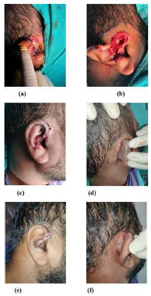
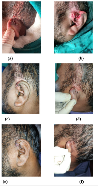
The patient was taken to the emergency operation theatre for cleaning of the wound and re-suturing. The right ear’s sutured wound was cleaned and painted with iodopovidone. Ring block was given with 2% lignocaine with adrenaline 1:100,000. Previous sutures were removed, and the lacerated wound was thoroughly cleaned with saline. All the foreign bodies were removed and devitalized, and dead tissues were removed. There was an incomplete right ear amputation present, and the ear remained attached by a 1.5 cm strip of skin at the level of tragus. The edges of the lacerated wound were then sutured using nylon 4-0 suture (Figure 1a-1f). After this left ear was cleaned, previous sutures were removed and the lacerated wound was thoroughly cleaned with saline and assessed. Dead tissue was removed. Through and through lacerated wound was present involving the root of the helix with exposure of cartilage reaching up tothe triangular fossa and some part of cymba concha. The ear wound was repaired by using nylon 4-0 sutures (Figure 2a-2f) and bilateral pressure dressing was done and the patient was kept on IV antibiotics with symptomatic treatment. Daily dressing was done for the next 7 days with the wound assessment. Sutures were removed after 7 days and the wound healed well with no residual necrosis. A good cosmetic result was achieved.
Figure 1:Right ear (a) sutured wound at the time of presentation. (b) wound after the removal of sutures and cleaning. (c) after resuturing with nylon 4-0 lateral view (d) posterior view (e) at postop day 7 after suture removal lateral view (f) posterior view.

Figure 2:Left Ear (a) sutured wound at the time of presentation. (b) wound after the removal of sutures and cleaning. (c) after resuturing with nylon 4-0 lateral view (d) posterior view (e) at postop day 7 after suture removal lateral view (f) posterior view.

Discussion
The pinna is formed from a single piece of elastic cartilage which on its lateral surface is highly adherent to the skin whereas slightly loose on its medial surface [3]. It gets its blood supply from branches of the external carotid artery which includes posterior auricular, temporal, occipital and maxillary (the deep auricular branch) arteries. Because of the protruded nature of elastic cartilage of the pinna, it is more prone to injuries due to trauma and thus post-traumatic infections [5].
Pinna lacerations are the most common injuries to the ear [6]. Most of them are caused by road traffic accidents. The goal of treatment is to maintain a balance between minimal debridement with tissue preservation as much as possible and to achieve the normal shape and structure without infection and thus prevent complications like haematoma formation and perichondritis of pinna. Haematoma formation in the sub perichondrial space can cause infection, necrosis, and new irregularly shaped cartilage formation which finally will lead to cauliflower ear [3].
Perichondritis is the most serious complication which leads to redness of the skin, pain, local temperature rise and finally the formation of an abscess in which immediate pus drainage is important to prevent the deformity [7]. Prevention of these complications can be achieved with early intervention, debridement of wound, meticulous suturing and proper antibiotic coverage [4].
As the ear is innervated by many nerve branches, a ring block can be given to provide adequate anaesthesia. Because of the superficial vasculature in this area, we should always aspirate before giving the injection. If the superficial temporal artery is punctured accidentally, application of the firm pressure should be done to prevent the formation of haematoma. The wound should then be washed thoroughly with saline to remove foreign bodies. Trimming of the irregular skin and removal of dead tissue should be done. The lacerated wound should be sutured meticulously with a nonabsorbable suture along with pressure dressing and prophylactic antibiotics to have proper healing without complications [8]. Regular follow-up of the patient should be done to look for the development of any complication.
Conclusion
Pinna lacerations are the most common injuries to the ear following mechanical trauma. Timely surgical intervention by thoroughly cleaning the wound, adequate debridement of dead tissue, trimming of irregular skin, meticulous suturing of lacerated wound, daily wound dressing with contour maintenance and wide antibiotic coverage is very important for healing without complications. Regular follow-up is required for early identification of any complication.
References
- Nojoumi A, Woo BM (2021) Management of ear trauma. Oral Maxillofac Surg Clin North Am 33(3): 305-315.
- Erdmann D, Bruno AD, Follmar KE, Stokes YH, Gonyon DL, et al. (2009) The helical arcade: Anatomic basis for survival in near-total ear avulsion. J Craniofac Surg 20: 245-248.
- Saimanohar S, Gadag RP, Subramaniam V (2012) A study of pinna injuries and their management. International Journal of Health and Rehabilitation Sciences 1(2): 81-86.
- Ahmad SSV, Mukundan A, Githin CR, Mary L (2017) Pinna injuries: Our experience. Journal of Medical Science and Clinical Research 5(3): 18782-18786.
- Menon A, Alagesan G (2018) Traumatic partial avulsion of pinna reconstruction with Limberg flap. World J Plast Surg 7(2): 231-234.
- Steele BD, Brennan PO (2002) A prospective survey of patients with presumed accidental ear injury presenting to a paediatric accident and emergency department. Emerg Med J 19(3): 226-228.
- Wright D (1987) Diseases of external ear. Scott Brown’s Otolaryngology (6th edn), Booth JB, Butterworth-Heinmann (Eds.), Oxford, England, 3: 1-20.
- Turpin IM (1990) Microsurgical replantation of the external ear. Clin Plast Surg 17(2): 397-404.
© 2024 Apoorva Kumar Pandey. This is an open access article distributed under the terms of the Creative Commons Attribution License , which permits unrestricted use, distribution, and build upon your work non-commercially.
 a Creative Commons Attribution 4.0 International License. Based on a work at www.crimsonpublishers.com.
Best viewed in


.jpg)


 Editorial Board Registrations
 Submit your Article
 Refer a Friend
 Advertise With Us
.jpg)


.jpg)


.bmp)
.jpg)
.png)
.jpg)


.jpg)


.png)

.png)


.png)


