- Submissions

Full Text
Experiments in Rhinology & Otolaryngology
A Rare Case of Cervical T.B Spine Causing Prevertebral Abscess & Recurrent Laryngeal Nerve Paralysis of Right Side
George MV*, Regi George AN and Divya Ambooken
Department of ENT, Jubilee Mission Medical College, India
*Corresponding author: George MV, Department of ENT, Head and Neck Surgery, Jubilee Mission Medical College, Thrissur, India
Submission: August 06, 2018;Published: September 21, 2018

ISSN 2637-7780 Volume2 Issue4
Introduction
Tuberculosis (TB) of the spine (Pott’s disease) is one of the most common and dangerous form of extra pulmonary TB infection. But Cervical spine tuberculosis is an uncommon form of tuberculosis of the spine. Usually lower thoracic spine is involved, followed by the middle thoracic and finally only the cervical spine gets involved with tuberculosis [1]. In 1779, Percivall Pott, for whom the disease is named, presented the classic description of spinal tuberculosis. TB of the cervical spine also called Morb Pott disease, or tuberculous spondylitis [2]. Spinal caries is the most common and is the most dangerous form of skeletal tuberculosis [2].
The common forms of presentation are constitutional symptoms like weakness, loss of appetite and weight evening rise of temperature and night sweating. Locally, there can be pain and stiffness of the neck [3,4]. The other form of manifestation is Neurological symptoms [5]. But our patient has presented in a different way.
Case Report
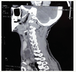
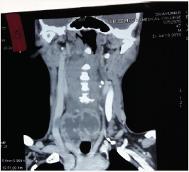
This is a case report of a 47 years old male patient who presented with right sided neck swelling and change in voice for 3 months. On examination, he was emaciated with sunken eyes and was weighing only 43 kilograms. There was a soft irregular swelling on right side of his neck. On indirect laryngoscopy, his right vocal cord was seen fixed. Chest X ray was taken which was almost normal. Blood picture showed elevated ESR, lymphocytosis. With this background, Patient was advised a CT scan of neck. CT neck showed prevertebral collection between trachea and carotid vessels on right side. Multiple enhancing lymph nodes also were seen and fibro cavitary changes involving posterior segment of left upper lobe and severe degenerative changes involving C4 to C7 vertebrae were noticed suggestive of prevertebral abscess with multiple necrotic lymph nodes. USG guided aspiration of around 7ml of pus was done from the abscess and sent for AFB staining. This was found to be positive for AFB (Figure 1). Then he was sent for an Orthopaedic consultation and they advised to use a cervical collar for restrictions of neck movements. Now he is on Anti Tubercular Therapy as advised by the Pulmonologist.
Figure 1:Sagittal reconstruction.

Discussion
Figure 2:Coronal reconstruction.

Usually, two contiguous vertebrae are involved but several vertebrae may be affected and skip lesions are also seen. The exudate penetrates the ligaments and follows the path of least resistance along fascial planes, blood vessels and nerves, to distant sites from the original bony lesion as cold abscess. In the cervical region, the exudate collects behind prevertebral fascia and may protrude forward as a retropharyngeal abscess Retropharyngeal abscess can present with local pressure effects such as dysphagia, dyspnoea, or hoarseness of voice (Figure 2). Further, dysphagia may also occur due to mediastinal abscess. Unlike retropharyngeal infection, cervical prevertebral space infection is most often caused by coagulase-positive staphylococci or Mycobacterium tuberculosis [6].
Only 10 case reports of tuberculosis retropharyngeal abscess had been appeared in the world literature from 1930 to 1973 [7]. Deep neck abscess may be life threatening because of the possibilities of airway obstruction, involvement of the carotid sheath, spread into the mediastinum, or septic shock. The mortality rate of patients with such life-threatening complications is as high as 40-50% [6]. Change of voice is possible in prevertebral abscess [1]. But an article on the causes of recurrent laryngeal nerve paralysis, doesn’t include this as a cause for the same, may be because it is extremely rare. This is after a study of 466 patients from October 1996 to December 1997 [8].
Conclusion
The report of this case about the vocal cord paralysis following a prevertebral abscess due to a Pott’s spine neck is a very rare presentation as the hoarse voice may not be due to a vocal cord paralysis but may be due to inflammation. Vocal cord paralysis produces a breathy voice rather than hoarseness.
References
- Sharma SK, Mohan A (2004) Extrapulmonary tuberculosis. Indian J Med Res 120(4): 316-353.
- Cristina OL, Gina E, Simona AD (2018) Tuberculosis of the cervical spine morb pott cervicalis our experience. Glob J Oto 13(3): 555861.
- Behari S, Nayak SR, Bhargava V, Banerji D, Chhabra DK, et al. (2003) Craniocervical tuberculosis: protocol of surgical management. Neurosurgery 52(1): 72-81.
- Hsu LC, Leong JC (1984) Tuberculosis of the lower cervical spine (C2 to C7). A report on 40 cases. J Bone Joint Surg Br 66(1): 1-5.
- Fang D, Leong JC, Fang HS (1983) Tuberculosis of the upper cervical spine. J Bone Joint Surg Br 65(1): 47-50.
- Herr RD, Murdock RT, Davis RK (1991) Serious soft tissue infections of the head and neck. Am Fam Physician 44(3): 878-892.
- Faidas A, Ferguson JV, Nelson JE, Baddour LM (1994) Cervical vertebral osteomyelitis presenting as a retropharyngeal abscess. Clin Infect Dis 18(6): 992-994.
- Myssiorek D (2004) Recurrent laryngeal nerve paralysis: anatomy and etiology. Otolaryngol Clin North Am 37(1): 25-44.
© 2018 George MV. This is an open access article distributed under the terms of the Creative Commons Attribution License , which permits unrestricted use, distribution, and build upon your work non-commercially.
 a Creative Commons Attribution 4.0 International License. Based on a work at www.crimsonpublishers.com.
Best viewed in


.jpg)


 Editorial Board Registrations
 Submit your Article
 Refer a Friend
 Advertise With Us
.jpg)


.jpg)


.bmp)
.jpg)
.png)
.jpg)


.jpg)


.png)

.png)


.png)


