- Submissions

Full Text
Experiments in Rhinology & Otolaryngology
A Rare Case of Nasal Inferior Meatus Polyps
George MV*
Professor of ENT Jubilee Mission Medical College Hospital, Kerala University of Health Sciences, India
*Corresponding author: George MV, Professor of ENT Jubilee Mission Medical College Hospital, Kerala University of Health Sciences, Thrissur, India
Submission: January 19, 2018;Published: April 10, 2018

ISSN: 2637-7780Volume1 Issue3
Abstract
This is a case report of a patient with a sided nasal inferior meatus polyposis. A 34-year-old female patient rested with history of discharge coming from right side of nose of 6 months duration. On examination, there was minimal discharge in the floor of the nose. On endoscopy, there was a swelling under the inferior turbinate. CT PNS showed an enlarged inferior turbinate of the same side and bilateral concha bullosa, and mild maxillary sinusitis. The patient had undergone an endoscopic excision biopsy of the swelling and the histopathological report was Inflammatory Nasal polyps.
Keywords: Post-thyroidectomy; Recurrent laryngeal nerve palsy; Arytenoidectomy
Introduction
A nasal polyp is considered as an inflammatory condition in nasal and paranasal sinus cavities and is frequently seen in all ENT clinics. The two usual types of polyps are the ethmoid polyps which are usually bilateral and the AC polyp which is often unilateral, both from the middle meatus. Polyps of the inferior meatus are not described.
Case Report
Our patient, a 34-year-old lady presented to our department with complaints of mild discharge coming from Right side of the nose of 6 months duration. She was not having any other symptoms related to nose like bleeding, headache, foul smell, or nasal obstruction. She never had any surgery inside her nose, or trauma, or foreign body inside the nose or history of packing the nose in the past.
Figure 1:

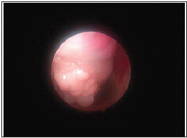
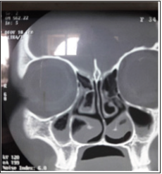
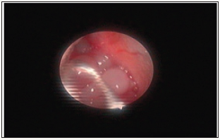
Her nasal cavity of Right side showed minimal discharge, and on endoscopy, an irregular bulge was seen on the under surface of inferior turbinate. There were no polyps in other areas like middle meatus or surroundings (Figure 1). CT scan showed a large inferior turbinate on the right side, maxillary sinusitis, and bilateral concha bullosa (Figure 2). Because of this atypical picture, she was taken up for surgery of excision biopsy of the mass (Figure 3). The histopathological examination revealed polypoid mucosa, lined by respiratory epithelium, sub epithelium showing marked oedema, and chronic inflammatory cell infiltration of lymphocytes and plasma cells.
Figure 2:

Figure 3:

Discussion and Review of Literature
Many pathogenic theories are proposed as etiology like adenoma and fibroma theory, glandular cyst theory, mucosal exudation block theory, glandular hyperplasia, gand new formation, ion transport theory, and likewise [1] most polyps originated from the clefts of the ostiomeatal complex region and the adjacent area. [2]. It is very rare to have polyp of inferior meatus. A Chinese author had described this in 2013 [3]. It was a female patient, 24 years old with a history of mass coming from the nose. On examination it was found out that the mass was coming from inferior meatus and going back. This is an apt description of nasal rhinosporidiosis. The rest of the cases reported were about choanal polyps arising from the inferior turbinate [4]. The literature doesn’t contain much about this rare condition.
Conclusion
This is a very rare condition and probably the 2nd case in world literature.
References
- Dagli M, Eryilmaz A, Besler T, Akmansu H, Acar A, et al. (2004) Role of free radicals and antioxidants in nasal polyps. The Laryngoscope 114(7): 1200-1203.
- Virat MD (2005) Update on nasal polyps: Etiopathogenesis. Journal Medical Association of Thailand 88(12): 1966-1972.
- Lei PS, Zheng Chunquan (2013) A case of inferior nasal polyps from the inferior turbinate. Chinese Journal of Otorhinolaryngology Head and Neck Surgery 48 (4): 299-299.
- Tuz M, Yasan H (2006) Choanal polyp originated from the inferior turbinate presenting as nasal polyposis. Kulak Burun BogazIhtis Derg 16(1): 37-40.
- Aydil U, Karadeniz H, Sahin C (2008) Choanal polyp originated from the inferior nasal concha. European Archives of Oto Rhino Laryngology 265(4): 477-479.
© 2018 George MV. This is an open access article distributed under the terms of the Creative Commons Attribution License , which permits unrestricted use, distribution, and build upon your work non-commercially.
 a Creative Commons Attribution 4.0 International License. Based on a work at www.crimsonpublishers.com.
Best viewed in


.jpg)


 Editorial Board Registrations
 Submit your Article
 Refer a Friend
 Advertise With Us
.jpg)


.jpg)


.bmp)
.jpg)
.png)
.jpg)


.jpg)


.png)

.png)


.png)


