- Submissions

Full Text
Examines in Physical Medicine and Rehabilitation: Open Access
Autonomic Dysreflexia, A Clinical Entity not to be Forgotten!
André V Borges*, Igor S Neto, Rodrigo Correia, Maria Cunha and Fátima Gandarez
Northern Rehabilitation Center, Portugal
*Corresponding author:André V Borges, Northern Rehabilitation Center, Portugal
Submission: August 5, 2022; Published: November 08, 2022

ISSN 2637-7934 Volume3 Issue5
Abstract
Introduction: Traumatic Spinal Cord Injury (TSCI) causes a constellation of interrelated autonomic
and cardiovascular abnormalities. One of them is Autonomic Dysreflexia (AD) which could appear when
the TSCI is at or above the sixth thoracic (T6) spinal cord segment, resulting from a parasympathetic
imbalance. It is clinically defined as acute hypertension generated by unmodulated sympathetic reflexes
below the injury level. In response to hypertension, the baroreflex system lowers blood pressure by
reducing heart rate and decreasing the activity of vasoconstrictor Sympathetic Preganglionic Neurons
(SPN) located in the thoracolumbar spinal cord. While vagal parasympathetic innervation of the heart
remains intact after TSCI, the disruption of descending vasomotor pathways to SPN produces an
incomplete compensatory decrease in peripheral vascular resistance so that hypertension persists until
the triggering stimulus is removed. The objective of this report is to describe a clinical case that suddenly
triggered AD after an uncommon cause, that of T2 AIS A (ASIA/ISCoS) (1) A in which AD harmed the life
of the patient but also the rehabilitation program.
Case report: A male 55 years with a prior history of TSCI, paraplegia AIS A, neurological level T2, was
admitted to the Northern Rehabilitation Center (NRC) for optimization of the neuromotor rehabilitation,
improving autonomy in daily life activities, and reducing joint limitations. The patient, after one weekend
moving home 3 days after that suddenly start with AD symptoms during the rehabilitation program,
triggered in the sitting position. Besides persistent hypertension, palpitations, diaphoresis, shivering, and
tachycardia. The patient was diagnosed after finding out from the clinical history that the cause of the AD
was a fracture of the left iliopubic and ischiopubic branches that would have arisen from a small accident
that occurred over the weekend patient had not even appreciated. The cause of AD is concluded after
being excluded from the most common AD triggers and conservatively treated.
Discussion: This cause draws attention even in low-energy injuries plus the prolonged immobilization
known in TSCI resulting in bone loss and osteoporosis could easily develop a fracture which could
trigger an AD and compromise all the rehabilitation programs, and the importance of anti-osteoporotic
treatment whenever is necessary. AD is a medical emergency that can put the patient at risk of life, so the
medical team should always be aware of this clinical entity.
Keywords: Dysreflexia; Parasympathetic imbalance; Clinical entity; Baroreflex system; Hypertension; Rehabilitation program
Introduction
In addition to the motor and sensory deficits, Traumatic Spinal Cord Injury (TSCI) causes a constellation of interrelated autonomic and cardiovascular abnormalities [1,2]. SCI at or above the sixth thoracic (T6) spinal cord segment often results in the development of Autonomic Dysreflexia (AD). AD is clinically defined as acute hypertension generated by unmodulated sympathetic reflexes below the injury level. In response to hypertension, the baroreflex system lowers blood pressure by reducing heart rate and decreasing the activity of vasoconstrictor Sympathetic Preganglionic Neurons (SPN) located in the thoracolumbar spinal cord. While vagal parasympathetic innervation of the heart remains intact after TSCI, the disruption of descending vasomotor pathways to SPN produces an incomplete compensatory decrease in peripheral vascular resistance so that hypertension persists until the triggering stimulus is removed. Severe cases that do not receive rapid treatment can have serious consequences such as stroke, cardiac arrest, seizure, and even death [3]. The most common triggers are over-distension of the bowel or bladder [4], other noxious stimuli including skin lacerations, ingrown toenails, pressure sores, tight clothing, and fractures. One of the highest priorities of the TSCI is mitigating AD and its trigger stimuli [5].
AD is most often present in the chronic phase of SCI, with a majority of cases first occurring 3-6 months after injury. It occurs in an emergency context and severe cases can cause death.
Case Presentation
Male, 55 years old, relevant personal history stand out for spinal cord injury 33 years after motorcycle accident resulting in AIS paraplegia A single neurological level T2 [1]. Since then, He is partially dependent on activities of daily living (self-care, food, self-propelled wheelchair, and adapted car driving). Until now, as complications, he only had various urinary tract infections (last in May 2018) and pressure ulcers in the buttock region (between 2013 and 2016). From surgical history had colostomy (since 2008), suprapubic cystostomy (since December 2017), and an osteosynthesis material from orthopaedic surgery (femur long nailing). He was admitted to the Northern Rehabilitation Center (NRC) in November 2018 to optimize neuromotor rehabilitation, improve autonomy in daily life activities, and reduce joint limitations. During the hospitalization period (December 2018 to January 2019), the rehabilitation program was harmed by the development of sudden episodes of autonomic dysreflexia, particularly in December. These episodes prevented the patient from performing the intensive rehabilitation program in the gyms and were triggered in the sitting position.
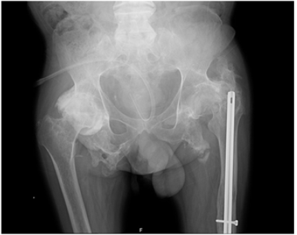
The medical team carried out the necessary studies to determine the triggering causes. From everything verified, the only relevant clinical information was a car accident that took place when the patient moved home for the therapeutic weekend. An x-ray was performed on 18-12-2019 at the NRC, which showed suspicion of a new recent fracture at the pelvic branch, not present in previous exams (Figure 1). The patient was referred and evaluated by orthopedics at the Vila Nova de Gaia Hospital Centre on 26-12-2018. Computed axial tomography was performed, showing marked osteoporosis, particularly in the sacrum region, and the presence of continuity solution involving the left ilio-pubic and ischium-pubic branches, without relevant bone deviations. After evaluation, it was reported that there was no need for specific care for this clinical situation, but radiological control of the pelvis should be performed and evaluated in an orthopaedic consultation. As a conclusion and after medical investigation, the most likely noxious stimulus was a fracture of the left iliopubic and ischiopubic branches.
Figure 1:Pelvis x-ray realized in 18-12-2019.

Discussion
Prolonged immobilization is known to result in bone loss and osteoporosis, TSCI is the maximum expression of this condition. Indeed, TSCI has been associated with a marked increase in bone loss below the TSCI level and a consequent increased risk of fractures. Osteoporosis may reach 61% of patients with TSCI [6]. These patients have an increased risk of skeletal fractures, ranging from 1 to 34%. Time since the injury, the severity, the level and aetiology of SCI, or the loss of bone mineral density, may partially explain the variability. Literature refers that anti-osteoporotic treatment in these patients is uncommon. Fractures in these patients present some findings, such as their relationship to lowenergy injuries, and the site, frequently located in bones distal the level of neurologic impairment, the femur distal and/or the tibia or fibula proximal [6,7]. This case describes a patient who developed an autonomic dysreflexia after an uncommon fracture (left iliopubic and ischium-pubic branches). As previously described, AD occurs in SCI after a noxious stimulus. Most patients with TSCI above T6 develop autonomic dysreflexia, and it is an entity that the medical rehabilitation team should be aware of. In this case, the medical team ruled out the most frequent triggers. After 3 days of research, the patient revealed that he was involved in a car accident 1 week ago. Fractures occur in a high frequency in active paraplegic patients after low-impact injuries during transfer or activities that involve minimal trauma [8,9].
Conclusion
AD is very common in these patients. It is a medical emergency that can put the patient at risk of life, so the medical team should always be aware of this clinical entity. The risk of fracture in these patients is high, especially below the level of spinal cord injury. In addition, a high percentage of osteoporosis is described and only a minority of patients undergo treatment. Is it important to medicate these patients? Will there be a statistically significant difference? Further studies need to be conducted taking these variables into account.
Appendices
Figure 1 Pelvis x-ray realized in 18-12-2019 shows extensive changes in the spine with osteophytes; Right coxarthrosis with shortening of the cervix; Status after left coxarthrosis with signs of old femoral fracture of the sub capital with an endomedullary rod; fracture of the ischiopubic rami bilaterally with heterotopic calcifications of the adductor muscles arising from the base of the branches (ossificans myositis). Degenerative alterations of the iliac sacrum.
References
- Kirshblum SC, Burns SP, Biering-Sorensen F, Donovan W, Graves DE, et al. (2011) International standards for neurological classification of spinal cord injury. J Spinal C 34(6): 535-546.
- Cragg JJ, Noonan VK, Krassioukov A, Borisoff J (2013) Cardiovascular disease and spinal cord injury: Results from a national population health survey. Neurology 81(8): 723-728.
- Karlsson AK (1999) Autonomic dysreflexia. Spinal Cord 37(6): 383-391.
- Canon S, Shera A, Phan NMH, Lapicz L, Scheidweiler T, et al. (2015) Autonomic dysreflexia during urodynamics in children and adolescents with spinal cord injury or severe neurologic disease. Journal Pediatric Urology 11(1): 32.
- Anderson KD (2004) Targeting recovery: Priorities of the spinal cord injury population. J Neurotrauma 21(10): 1371-1383.
- Maimoun L, Fattal C, Micallef J-P, Peruchon E, Rabischong P (2006) Bone loss in spinal cord injured patients: From physiopatholy to therapy. Spinal Cord 44(4): 203-210.
- Gifre L, Joan Vidal, Josep Carrasco, Enric Portell, Josep Puig, et al. (2014) Incidence of skeletal fractures after traumatic spinal cord injury: 10-year follow-up study. Clinical Reabil 28(4): 361-369.
- Comarr AE, Hutchinson RH, Bors E (1962) Extremity fractures of patients with spinal cord injuries. Am J Surg 103: 732-739.
- Garland DE, Adkins RH, Kushwaha V, Stewart C (2004) Risk factors for osteoporosis at the knee in the spinal cord injury population. J Spinal Cord Med 27(3): 202-206.
© 2022 André V Borges. This is an open access article distributed under the terms of the Creative Commons Attribution License , which permits unrestricted use, distribution, and build upon your work non-commercially.
 a Creative Commons Attribution 4.0 International License. Based on a work at www.crimsonpublishers.com.
Best viewed in


.jpg)


 Editorial Board Registrations
 Submit your Article
 Refer a Friend
 Advertise With Us
.jpg)


.jpg)


.bmp)
.jpg)
.png)
.jpg)


.jpg)


.png)

.png)


.png)


