- Submissions

Full Text
Developments in Anaesthetics & Pain Management
Ultrasound-Guided Anesthetic Injections in Tennis Elbow-New Findings With Implications for Treatment
Möllestam K1,2 and Alfredson H3,4*
1Department of Clinical Sciences Lund-Orthopedics, Lund University, Sweden
2Department of Orthopedics, Hässleholm-Kristianstad Hospitals, Sweden
3Department of Community Medicine and Rehabilitation, Sports Medicine, Umeå University, Sweden
4Alfredson Tendon Clinic, Capio Ortho Center Skåne, Malmö, Sweden
*Corresponding author:Alfredson Hakan, Department of Community Medicine and Rehabilitation, Sports Medicine, Umeå University, Umeå and Alfredson Tendon Clinic, Capio Ortho Center Skåne, Malmö, Sweden
Submission:December 19, 2023;Published: January 12, 2024

ISSN: 2640-9399 Volume2 Issue5
Abstract
Background: Treatment of chronic painful tennis elbow is known to be difficult, and the source of
pain has not been clarified. A recent study has shown that the fibrous layer on the superficial side of
the extensor origin is rich in sensory nerves. This study aimed to evaluate the effects on grip pain and
strength after targeted injections of a local anesthetic, in patients with chronic painful Tennis elbow.
Material: Fourteen patients (6 men, mean age 60 years and 8 women, mean age 43 years) with clinical
and US-verified changes corresponding to the diagnosis Tennis Elbow in 15 elbows, were included. All
had had a long duration (average 19 months, range 6-48 months) of pain symptoms and undergone
multiple different treatments without effect.
Methods: A US-guided injection of 1-1.5ml Polidocanol (10mg/ml) was placed targeting the region with
high blood flow in the fibrous tissue layer on the superficial side of the extensor origin. Grip pain and
strength during a standardized dynamometer test was evaluated on a 10cm VAS scale, before and 15
minutes after injection.
Result: In 12/14 patients (13/15 elbows) there was decreased pain during gripping (VAS from mean 7
(SD 1.3) to mean 2.5 (SD 1.6), p<0.05), and increased strength during straight arm test (from mean 30kg
(SD 10.7) to mean 39kg (SD 13.5), p<0.05).
Conclusion: In patients with Tennis elbow, there were significant effects on pain and grip strength after
US-guided injections of a local anesthetic targeting the nerve rich fibrous layer on the superficial side of
the extensor origin. These findings might have implications for treatment.
Keywords:Tennis elbow; Anesthetic injections; Straight arm test; Sensory nerves; Achilles; Patellar tendinopathy
Introduction
Tennis elbow is a non-inflammatory condition, also called lateral elbow tendinopathy, with unknown aetiology and pathogenesis [1-3]. The origin to pain is not known, but the general opinion is that the Extensor Carpi Radialis Brevis (ECRB) muscle and its tendinous origin plays a central role [4-6]. Tennis elbow is a condition well known to be troublesome to treat [7]. First, non-invasive treatments like NSAIDs and physiotherapy [8], orthotic devices [9] and rest, are being used. Secondly, different types of injections [10-14] are tried, and finally, surgical treatments [15] might be indicated. The results after treatment are varying and not convincing, and for this chronic painful condition there is a general opinion that there is no golden standard treatment.
The pain mechanisms related to Tennis elbow have not been fully clarified, and there is a need for studies evaluating where the pain comes from. For Achilles and patellar tendinopathy, studies have shown that the majority of sensory nerves are located outside the tendons [16,17], and a recent study on patients with chronic painful Tennis elbow Ultrasound (US) and Colour Doppler (CD)-guided biopsies showed that the superficial fibrous tissue was richly innervated by sensory nerves [18]. The aim with the current study was to study the effects on pain and grip strength after targeted US and CD-guided injections of a local anesthetic in patients with chronic painful Tennis elbow.
Material and Methods
Participants
Fourteen patients (8 women and 6 men, mean age 50 years, range 35 to 70 years) with a long duration of pain (mean 19 months, range 6 to 48 months) diagnosed as tennis elbow, in altogether 15 elbows (13 unilateral, 1 bilateral), were included. All had undergone multiple different treatments such as physiotherapy including stretching exercises (n=12), eccentric exercise (n=10), medication with NSAIDs (n=9), tennis elbow brace (n=9), acupuncture (n=7), shock wave (n=3), and cortisone injections (n=9). The clinical diagnosis tennis elbow was based on pain on palpation of the extensor origin, pain during resisted wrist extension, and pain during resisted third finger test. Patients with differential diagnoses such as cervico-brachialgia, radial nerve compression, previous operations in the elbow, chronic inflammatory conditions, and restricted elbow ROM were excluded based on history and clinical findings.
Ultrasound (US) and colour doppler (CD) examination
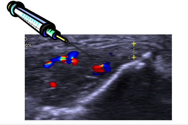
All tendons were examined with high resolution grey scale- Ultrasound (US) and Colour Doppler (CD), Acuson Sequoia 512, with 8-13MHz frequency. The examinations were carried out in a sitting position, with the arm resting on a table, having the elbow flexed to 70-80 degrees and the wrist in neutral position. The same experienced Doctor (H.A.) performed all US-and CD-examinations. In all patients there were structural abnormalities (including hypoechoic regions, irregular structure, or calcifications) in the extensor origin and a bone spur of varying size on the lateral epicondyle (lateral epicondyle enthesophyte) (Figure 1). In all patients there was a thick fibrous layer including high blood flow (with vessels entering the underlaying origin) on the superficial side of the extensor origin (Figure 1).
Figure 1:Grey scale and colour doppler ultrasound picture of the extensor origin in a patient with chronic painful Tennis elbow. There are structural changes including hypo-echoic regions in the extensor origin, and a thick fibrous layer (marked) including high blood flow on the superficial side of the extensor origin. There is a bone spur on the lateral epicondyle. The injection needle is positioned inside the thickened fibrous tissue, close to the regions with high blood flow, on the superficial side of the extensor origin.

Injection procedure
The active substance in Polidocanol is an aliphatic non-ionized nitrogen-free surface anesthetic. Before the treatment, the skin was washed with a solution of chlorhexidine and alcohol. The patients were sitting with their arms resting on a table, having 70-80 degrees of elbow flexion, and the wrist in neutral position. The US and CD-guided dynamic injections were performed with a 0.7x50mm needle, connected to a 2ml syringe, and the Polidocanol (10mg/ ml) injections targeted the regions with high blood flow (blood vessels) in the fibrous layer on the superficial side of the extensor origin only. The ultrasound probe was positioned longitudinally at the lateral epicondyle and along the origin, parallel with the fibers.
When the tip of the needle was positioned correctly, a small amount of Polidocanol was injected in fractions until all vessels were closed. Altogether 1-1.5ml was injected. It was possible to observe the immediate effect of the injection. If the position of the needle was correct (inside or very close to the vessels) the circulation in the target vessels stopped instantly.
Grip strength tests
Maximum voluntary grip strength was evaluated by using a hydraulic hand dynamometer (FEI Irvington, NY, USA). Maximum grip strength was measured three times, and the mean value was used for the statistical analysis. During the dynamometer-test, the arm was held in the horizontal plane, with the elbow straight, and the wrist in neutral position. The grip strength was measured before and 15 minutes after injection of Polidocanol.
Pain during gripping
Pain during the maximum grip test was recorded on a VAS scale. Using a 10cm Visual Analogue Scale (VAS) for pain (where no pain is recorded as 0 and severe pain as 10), the patient recorded the amount of elbow pain when gripping. Pain during gripping was evaluated both before and 15 minutes after injection of Polidocanol.
Statistical analysis
A paired t-test was used to compare the mean grip strength and pain during gripping before and after injection.
Ethics
There was ethical approval for this study, Dnr: 2010/170-31, from the Ethical Committee of the Medical Faculty, University of Umeå.
Result
In 12 of 14 patients (13/15 elbows) there was a significantly decreased pain during gripping from mean VAS 7 (range 5-9) to mean VAS 2.5 (range 1-6) (p<0.05). In the same 12 out of 14 patients there was a significantly increased muscle strength during straight arm test from 30kg (range 6-52) to 39kg (range 23-71) (p<0.05). In 2 patients (2 elbows) (VAS 8 and 7 respectively) there was no decreased pain during gripping and no increased muscle strength (11 and 23kg respectively) during strength test.
Discussion
The main finding in the current study was that US and CD-guided injections of a local anesthetic targeting the nerve rich fibrous tissue on the superficial side of the extensor origin decreased the pain and increased the muscle strength in patients suffering from chronic painful Tennis elbow. The patients represent a normal population first treated by general practitioners and physiotherapists, and then referred to the same county hospital (Hässleholm) in the south of Sweden, for treatment of Tennis elbow. All patients included in the current study had had a long duration of pain symptoms, and had tried the commonly used methods for treatment without success. Many had been treated with multiple cortisone injections into the extensor origin, one patient had been injected 8 times with cortisone, and were considered very difficult cases. The ultrasound examination used for diagnosis and inclusion showed that in more than 50% of the patients, there were major structural changes and a suspicion of partial ruptures in the extensor origin.
Previous studies using injection treatments have focused on injections placed inside the extensor origin, some with short term success [10-14], but no studies have shown good long term results. Regarding surgical treatments there is the same experience with often only short term success [15], and there is no golden standard method. Altogether, this indicates that the main location for pain has still not been identified. A recent study on US and CD-guided biopsies taken from the thick fibrous layer on the superficial side of the extensor origin in patients with chronic painful Tennis elbow provided new information about the location of nerves of possible importance for this condition [18]. Interestingly, this thick fibrous layer contained multiple sensory nerves, often located in close relation to blood vessels. This is, to the best of our knowledge, the first time the neural patterns on the outside of the extensor origin have been identified. Comparing with other tendinopathies, like Achilles and patellar tendinopathy, immunohistochemical analyses of biopsies showed that the majority of the sensory nerves involved in these conditions were found outside the tendons [16,17]. Over time, those findings have led to new successful mini-invasive treatments [19,20].
Previous studies using injections of Polidocanol targeting the inside of the extensor origin designed as a treatment method have shown temporary decreased pain and increased grip strength [14,21], and the conclusions from those studies where that the origin for pain was inside the extensor origin indicating that treatments should be focused on the inside. However, the longer term results were not convincing, and the results from the current study indicate that a major part of the pain has its source in the fibrous layer outside the superficial side of the extensor origin. The new findings from the current study opens up a new possible target for treatment.
We used VAS for pain during maximum gripping. VAS is known to be a reliable instrument and valid measure of pain intensity [22]. We also evaluated grip strength that is considered a good objective outcome measure for Tennis elbow [23]. We used US and CD examinations to evaluate the extensor origin and surrounding tissues. There are some limitations using greyscale ultrasonography and especially Doppler examinations. The method is examiner dependant [24]. There is no reliable method to calculate the flow. Positioning and pressure on the probe will affect the findings [24]. The examination is also depending on the patient, if tension in the wrist extensors is high, due to muscle contraction or by palmar flexion of the wrist, the blood flow is affected (decreased). Therefore, in the current study we used an experienced doctor (H.A.) for all US and CD examinations.
Ultrasound-guided injections for treatment of Tennis elbow have also been also by other groups [25,26]. In those studies the injections were not targeted to the specific nerve rich tissue on the superficial side of the extensor origin, but instead most often placed inside the extensor origin. Also, ultrasound-guided needling (needle puncture treatment) of the extensor origin, where the needle is placed inside the extensor origin, has been tried for treatment of this condition [27].
It is of obvious importance to try to find better treatments for patients suffering from chronic painful Tennis elbow. We believe that the results of our study are of interest and should be followed by treatment studies targeting only the nerve rich fibrous layer on the superficial side of the extensor origin.
Conclusion
In conclusion, US and CD-guided injections targeting only the nerve rich fibrous tissue on the superficial side of the extensor origin temporarily decreased pain and increased muscle strength in patients with chronic painful therapy resistant Tennis elbow. The findings might have implications for targeted treatment of this condition.
References
- Boyer MI, Hastings H (1999) Lateral tennis elbow: Is there any science out there? J Shoulder Elbow Surg 8(5): 481-491.
- Alfredson H, Ljung BO, Thorsen K, Lorentzon R (2000) In vivo investigation of ECRB tendons with microdialysis technique--no signs of inflammation but high amounts of glutamate in tennis elbow. Acta orthopaedica Scandinavica 71(5): 475-479.
- Khan KM, Cook JL, Kannus P, Maffulli N, Bonar SF (2002) Time to abandon the "tendinitis" myth. BMJ 324(7338): 626-627.
- Nirschl RP (1992) Elbow tendinosis/tennis elbow. Clin Sports Med 11(4): 851-870.
- Regan W, Wold LE, Coonrad R, Morrey BF (1992) Microscopic histopathology of chronic refractory lateral epicondylitis. Am J Sports Med 20(6): 746-749.
- Potter HG, Hannafin JA, Morwessel RM, DiCarlo EF, O'Brien SJ, et al. (1995) Lateral epicondylitis: correlation of MR imaging, surgical, and histopathologic findings. Radiology 196(1): 43-46.
- Bisset L, Paungmali A, Vicenzino B, Beller E (2005) A systematic review and meta-analysis of clinical trials on physical interventions for lateral epicondylalgia. Br J Sports Med 39(7): 411-422.
- Green S, Buchbinder R, Barnsley L, Hall S, White M, et al. (2002) Non-steroidal anti-inflammatory drugs (NSAIDs) for treating lateral elbow pain in adults. Cochrane Database Syst Rev 2013(5): CD003686.
- Struijs PA, Smidt N, Arola H, Dijk VC, Buchbinder R, et al. (2002) Orthotic devices for the treatment of tennis elbow. Cochrane Database Syst Rev 1: CD001821.
- Smidt N, Assendelft WJ, van der Windt DA, Hay EM, Buchbinder R, et al. (2002) Corticosteroid injections for lateral epicondylitis: A systematic review. Pain 96(1-2): 23-40.
- Connell DA, Ali KE, Ahmad M, Lambert S, Corbett S, et al. (2006) Ultrasound-guided autologous blood injection for tennis elbow. Skeletal Radiol 35(6): 371-377.
- Mishra A, Pavelko T (2006) Treatment of chronic elbow tendinosis with buffered platelet-rich plasma. Am J Sports Med 34(11): 1774-1778.
- Placzek R, Drescher W, Deuretzbacher G, Hempfing A, Meiss AL (2007) Treatment of chronic radial epicondylitis with botulinum toxin A. A double-blind, placebo-controlled, randomized multicenter study. J Bone Joint Surg Am 89(2): 255-260.
- Zeisig E, Ohberg L, Alfredson H (2006) Sclerosing polidocanol injections in chronic painful tennis elbow-promising results in a pilot study. Knee Surg Sports Traumatol Arthrosc 14(11): 1218-1224.
- Buchbinder R, Johnston RV, Barnsley L, Assendelft WJ, Bell SN, et al. (2011) Surgery for lateral elbow pain. Cochrane Database Syst Rev 2011(3): CD003525.
- Andersson G, Danielson P, Alfredson H, Forsgren S (2007) Nerve-related characteristics of ventral paratendinous tissue in chronic Achilles tendinosis. Knee Surg Sports Traumatol Arthrosc 15(10): 1272-1279.
- Danielson P, Andersson G, Alfredson H, Forsgren S (2008) Marked sympathetic component in the perivascular innervation of the dorsal paratendinous tissue of the patellar tendon in arthroscopically treated tendinosis patients. Knee Surg Sports Traumatol Arthrosc 16(6): 621-626.
- Spang C, Alfredson H (2017) Richly innervated soft tissues covering the superficial aspect of the extensor origin in patients with chronic painful tennis elbow-Implication for treatment? J Musculoskelet Neuronal Interact 17(2): 97-103.
- Alfredson H (2011) Ultrasound and doppler-guided mini-surgery to treat midportion Achilles tendinosis: Results of a large material and a randomised study comparing two scraping techniques. Br J Sports Med 45(5): 407-410.
- Masci L, Alfredson H, Neal B, Bee WW (2020) Ultrasound-guided tendon debridement improves pain, function and structure in persistent patellar tendinopathy: Short term follow-up of a case series. BMJ Open Sport Exerc Med 6(1): e000803.
- Zeisig E, Fahlström M, Öhberg L, Alfredson H (2008) Pain relief after intratendinous injections in patients with tennis elbow: Results of a randomised study. Br J Sports Med 42(4): 267-271.
- Thong ISK, Jensen MP, Miró J, Tan G (2018) The validity of pain intensity measures: what do the NRS, VAS, VRS, and FPS-R measure? Scand J Pain 18(1): 99-107.
- Bateman M, Evans JP, Vuvan V, Jones V, Watts AC, et al. (2022) Development of a core outcome set for lateral elbow tendinopathy (COS-LET) using best available evidence and an international consensus process. Br J Sports Med 56(12): 657-666.
- Levin D, Nazarian LN, Miller TT, Kane PLO, Feld RI, et al. (2005) Lateral epicondylitis of the elbow: US findings. Radiology 237(1): 230-234.
- Mezian K, Jačisko J, Novotný T, Hrehová L, Angerová Y, et al. (2021) Ultrasound-guided procedures in common tendinopathies at the elbow: From image to needle. Appl Sci 11(8): 3431.
- Ricci V, Mezian K, Cocco G, Tamborrini G, Fari G, et al. (2023) Ultrasonography for injecting (around) the lateral epicondyle: EURO-MUSCULUS/USPRM perspective. Diagnostics (Basel) 13(4): 717.
- Zhu J, Hu B, Xing C, Li J (2008) Ultrasound-guided, minimally invasive, percutaneous needle puncture treatment for tennis elbow. Adv Ther 25(10): 1031-1036.
© 2024 Alfredson H. This is an open access article distributed under the terms of the Creative Commons Attribution License , which permits unrestricted use, distribution, and build upon your work non-commercially.
 a Creative Commons Attribution 4.0 International License. Based on a work at www.crimsonpublishers.com.
Best viewed in


.jpg)


 Editorial Board Registrations
 Submit your Article
 Refer a Friend
 Advertise With Us
.jpg)


.jpg)


.bmp)
.jpg)
.png)
.jpg)


.jpg)


.png)

.png)


.png)


