- Submissions

Full Text
Clinical Research in Animal Science
Joint Laxity in Clinical Normal Dogs
Jens Arnbjerg*
Department of Veterinary Diagnostic Imaging Sund, University of Copenhagen, Denmark
*Corresponding author: Jens Arnbjerg, Department of Veterinary Diagnostic Imaging Sund, University of Copenhagen, Dyrlaegevej 32, 1870 Frederiksbergc, Denmark
Submission: August 06, 2021;Published: August 18, 2021

ISSN: 2770-6729Volume 1 - Issue 5
Abstract
Significance and Impact of the Study
It has earlier been described how clinical normal small breed dogs can have different joint laxity without this laxity leading to osteoarthrosis in the hip joints (HD).
These case reports show joint laxity in other joints than the hips in different breeds and even in larger breed dogs. The pictures are all taking without stress put on the joints by purpose, and most of the dogs not being clinical lame. Most of the cases did not show permanent clinical sign in relation to the joints examined.
Therefore, if there are no specific osteoarthritic finding in the joint, the laxity could be normal for this dog and the prognosis should not be evaluated so guarded as they often are just because of distance between joint elements. The parameters for screening programs should be more breed adapted and not just the same for all different breeds. Joint laxity is not always a warning of an osteoarthritis development.
In the international literature it is often stated a lack of agreement between the radiological evaluation and clinical picture which could be due to incorrect radiological interpretation
Keywords: Joint laxity; Osteoarthrosis; Breed depending joint laxity; Breed depending on parameters for HD; Clinical importance of Joint laxity
Introduction
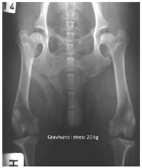
My interest in laxity in the joint was established some 30-35 years ago when a student wanted to have her Dachshund ´s hip examined. It was in the days when we did not tranquilize the dogs before taking the hips, so we retained the dog manually. We heard a small “klik”- X-ray pictures were taken, and the picture was similar to Figure 1. The dog was running around like nothing had happened-so we did not warry too much. The dog did not develop any side effect 5 years after the “accident”.
Figure 1: This is a radiograph of a dachshund where the hindlegs are pulled distally with 20kg (measured with by a spring-balance). The femoral heads are dislocated distally, and air-bubbles are noticed within the joints (vacuum phenomenon). After the radiograph was taken the dog was euthanized due to other reasons and the joints were opened. The joints were normal-no joint elements were damaged.
The purpose of the study was to draw the attention to some physiological breed depending on laxity in the joints, so one do not over-interpret a joint laxity, which in some other breeds can play a role in developing osteoarthrosis e.g. Labrador and German shepherd.
Material and Methods
The material consists of different dogs radiographed for different diagnostic reasons mainly without stress put on the region of interest by purpose presented in 6 different clinical cases.
Cases
Case 1
The dachshund dog shown in Figure 1, is from a project where 34 different dogs and breeds (under 15kg) where radiographs of the hips taken under stress where the hip were stressed by pulling exactly 20kg in the hindlegs (measured by a spring-balance). The study was published: Hip Joint Laxity in Small Dog Breeds [1].
Case 2
Figure 2:Left shoulder-joint of a golden retriever. Negative air-contrast of the joint was performed by inflation of 5 cc air. The bony elements of the Scapula-Humeral joint are separated in a significant way (2-3mm) due to the air-insufflation.
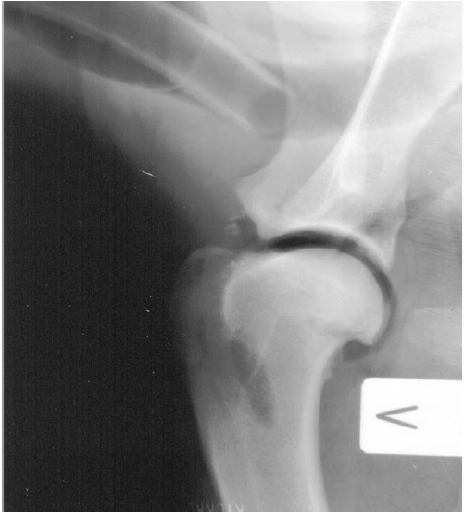
Before 1980-before ultrasound examinations were used in general, we often made contrast studies of joints by inflation of air into the joint space to visualize defects e.g., in the cartilage or joint mouses. Even under quite low pressure we produced a distance between the components of the joint and thereby visualizing their surfaces as it is seen in Figure 2. Only 5-10cc was injected into the joint space as in this case where the separation of the elements is some mms.
Case 3
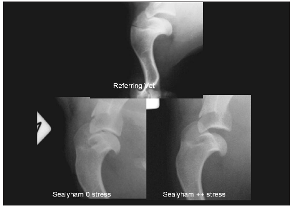
Figure 3 was a case where a 10-month-old Sealyham Terrierpuppy was radiographed because of lameness in the front leg. The practitioner foresaw a dramatic evolution of osteoarthrosis due to the open joint space in the shoulder joint and therefore recommended euthanasia of the dog. The breeder did not accept that proposal and therefore send it to a second opinion examination. The 2 lower pictures were taken-the one with and the other without stress in the front leg. The dog lived happy another 10 year.
Figure 3: A Sealyham Terrier - puppy, 10 months old - left shoulder region. The upper picture was taken at the referring vet’s clinic. A significant joint laxity is observed with a great opening at the joint caudally. The 2 lower pictures are taken at the Veterinary University Hospital. The left picture was taken with no stress and the right one with some stress by pulling the front leg cranially-a projection often seen in screening programs for OCD in the shoulder joint.
Case 4
Figure 4:Right shoulder of a 12-month-old, Old English Bulldog taken in a screening program for OCD in the shoulder joint. 2 arrows over the Tuber Scapula pointing on 2 air-bubbles in the shoulder joint. The dog had no clinical symptoms before or after the picture was taken.
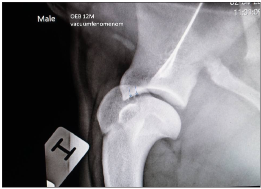
Figure 4A:An Old English Bulldog, 18 moth old. The pictures were sent for screening for OCD in the shoulder joints. The upper picture showing the right shoulder in the typical extending projection producing a shoulderjoint- picture outside the chest. The lower pictures shows the left front leg with the leg in a flexed projection, and no stress in the shoulder joint but the shoulder joint is not extended and therefore not so well presented superimposed over the more dense soft tissue.
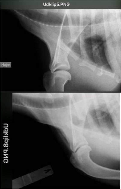
When screening for OCD in the shoulder joints incidentally we often observe a caudal opening of the shoulder joint just by pulling the leg cranially and dorsally to have the joint superimposed over the trachea as seen in Figure 4. In this case we can observe 2 small round dark air bubbles between scapula and humerus. These bubbles represent the vacuum-phenomenon arising from the sudden unset of under-pressure in the joint and thereby freed the solvable oxygen and carbon dioxide from the synovia. When that happens a small sound often can be heard. The same sound can often be heard when young people try to “break” their fingers. This radiograph was made as a routine screening picture taken of the front leg of this Old English Bulldog.
Case 5
Figure 4A This radiograph was taken of an 18-month-old Old English Bulldog taken as a part of a screening program for OCD in the shoulder. In the published paper [1] there was only one Old English Bulldog, but since then a bigger number of this breed has shown a number of joint laxity even it is a larger dog than the original material (<15kg).
Case 6
Figure 5 & 6 are radiographs from the same Staffordshire Bull Terrier taken under stress (Figure 5) so there is some distance between the acetabulum and the femoral heads. The referring vet wondered why the joint was so open and therefore he was afraid that he had destroyed something within the joint-even the dog was walking normally after the picture was taken.
Another picture was taken under more gentle circumstances (Figure 6). This is another example of a bigger dog with joint laxity taken incidentally.
The puppy had no clinical symptoms during the next 10 years.
Figure 5:Radiograph of the hip joints where there is quite a distance between acetabulum and the femoral heads due to pulling the legs distally. The radiographs are taken for evaluation of the hips for screening for HD (Staffordshire Bull Terrier) The femoral heads are dislocated 3-4mm distally. The same dog was presented too with radiographs taken with no stress in Figure 6.
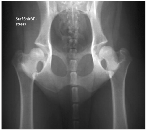
Figure 5:The same Staffordshire Bull Terrier as seen in Figure 5-but without pulling the legs during exposure.

Discussion
It has for quite a number of years-more than 50-been accepted that a number of orthopedic problems e.g., HD and AD has a hereditary reason. However, studies of the DNA in different breeds have shown that more than 25 loci are involved in HD [2] and therefore it is very difficult to produce a genetic depending on eradication programs to diminish the problem.
All the shown pictures except the contrast study of the scapulahumeral joint can be from daily life pictures in general practice and/or in different screening programs. So, no specific stress was by purpose put on the dogs during radiograph procedures. The traditional idea is that joint laxity results in osteoarthrosis in larger dog breeds, but these pictures show that in some breeds it is certainly not always the case - especially in smaller dog breeds. But not all small dog breeds have laxity in their joints. Sounds from the joints is not always a clear sign of malignancy as described in case 4 and by Arnbjerg [1]. In a number of orthopedic diseases pulling with quite large forces has been used as a therapeutic method, involving the joints as well as the muscles and by some veterinarians too [3].
In some breeds like Labrador Retriever, the subluxation might play a greater importance in developing osteoarthrosis later in life [4]. However, in small dog breeds some of them do not develop osteoarthrosis even they have severe joint laxity [1,5]. Therefor screening programs should use breed-depending parameters and not use the same parameters in all joints and breeds.
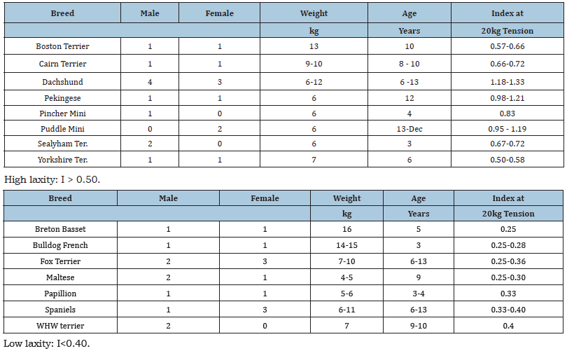
In small dog breeds there is a highly breed depending joint laxity, which is more specific depending to the breed and not to the individual dogs [1]; Table 1 & 2. However, lately several examples of larger dog breed dogs have been found incidentally, so it is probably not only some of the small dog breeds which can “suffer” from laxity in the joints in clinical healthy dogs.
Table 1: Different small dog breeds arranged according to joint-laxity-index and the standard deviation within the breed in 34 individual dogs. Generally, there are less variations within the breed than between some breeds.
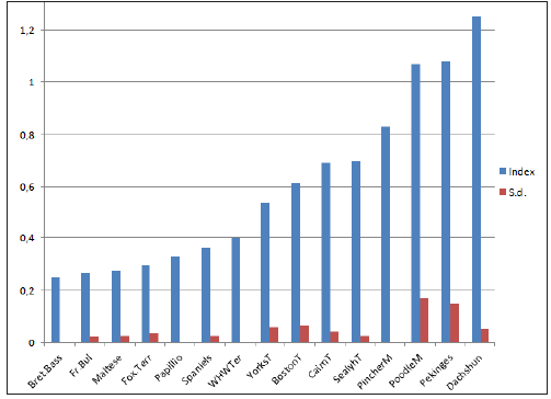
Table 2: Displacement-Index arranged according to high and low laxity in the hip joints in small dog breeds (<15kg).
Therefor some authors recommend different parameters used in evaluating joints in different breeds [1,2,6,7]. Many of the scores in the screening programs do not always show clinical signs matching the bad scores later in life, and the explanation can be a joint laxity which can be normal for the specific breeds and therefore often evaluated too badly. The same results are observed in a study of Sari et al. [8], where only one out of the 30 English Bulldogs (2-5 years old) were evaluated to have B. The rest had C, D, or E and 55% had E according to OFA scoring evaluation for HD, but only 6 showed pain response to palpation of the hip joints and only 4 showed signs of Osteoarthritis on the radiographs. The same tendency was found in the small dog breed Cairn Terrier [5]. So many individuals are taken out of breeding on false grounds.
Generally, joints with osteoarthrosis are tighter and show less joint laxity when exposed to stress. Therefore, if a joint shows osteoarthrosis the” normal” joint laxity for this specific breed are not expected.
Examination over longer periods in human patients [1,9] with daily stressing the joints with or without making noise, did not seem to have any damage to the joints. Therefore, the prognosis in cases with joint laxity should not be so guarded as previously given, unless specific clinical examination show sign of pain during manipulation on the joint involved and these joints are showing osteoarthritis. Making the same projection in the contralateral joint can also be an indicator of “normality” in a specific dog.
References
- Arnbjerg J (2017) Hip joint laxity in small dog breeds-A radiological study. SOJ Vet Sci 3(1): 1-5.
- Mainz E, Tellhelm B, Krawczk M (2017) Prospective evaluation of a patented DNA test for canine hip dysplasia. Plos One 12(8): e0182093.
- Arvidsen I (1990) The hip joint: forces needed for Distraction and appearance of the vacuum phenomenon. Scan J Rehab Med 22(3): 157-161.
- Smith GK, Lawler DF, Powers MY, Biery DN, Shofer F, et al. (2012) Chronology of hip dysplasia development in a cohort of 48 Labrador Retrievers followed for life. Vet Surg 41(1): 20-33.
- Arnbjerg J (2017) The prevalence of coxofemoral osteoarthritis in small dog breeds and HD screening in cairn terriers a clinical radiological study. SOJ Vet Sci 3(3): 1-5.
- Beuchat C (2017) Hip laxity and the risk of degenerative joint disease. Institute of Canine Bioloy.
- Read RA (2011) Carpal laxity syndrome in puppies. WSAVA proc.
- Mölsä SH, Hyytiäinen HK, Morelius KM, Palmu MK, Personen TS, et al. (2020) Radiographic findings have an association with weight bearing and locomotion in English bulldogs. Acta Vet Scand 62(1): 19.
- Protopas MG, Cymet TC (2002) Joint cracking and popping: Understanding noises that accompany articular release. J Am Osteopath Assoc 102(5): 283-287.
© 2021 Jens Arnbjerg. This is an open access article distributed under the terms of the Creative Commons Attribution License , which permits unrestricted use, distribution, and build upon your work non-commercially.
 a Creative Commons Attribution 4.0 International License. Based on a work at www.crimsonpublishers.com.
Best viewed in


.jpg)


 Editorial Board Registrations
 Submit your Article
 Refer a Friend
 Advertise With Us
.jpg)


.jpg)


.bmp)
.jpg)
.png)
.jpg)


.jpg)


.png)

.png)


.png)


