- Submissions

Full Text
COJ Technical & Scientific Research
DNA Typing: A Scientific Tool to Exonerate Innocent from Guilty
Praveesh Bhati* and Anil Kumar Singh
Department of Computer Science and Engineering, Nepal
*Corresponding author:Praveesh Bhati, DNA Finger Printing Unit, State Forensic Science Laboratory, India
Submission: June 6, 2019; Published: June 18, 2019

Volume2 Issue2June, 2019
Abstract
The accessibility of DNA examination has significantly altered the approach that offenses are scrutinized. The significance of outcome becomes more valuable when we pointed connection of individual to particular crime. DNA results can link offender to their crimes, exonerate wrongfully accused individuals, identify mass fatality victims and more. In the case of sexual assault when victim raised finger to the particular person that he was committed crime with her then scientific examination play important role. Microscopic examination that seeks for detection of spermatozoa alone cannot prove that a sexual assault has occurred by same suspect alleged by victim. DNA analysis can reveal whether a suspect’s DNA is, or is not, present on the source of victim. In present case study, the authors tried to give information to the readers with an overview of the limitations of traditional method for examination of sexual assault case in wrongful conviction of the suspect and also advantage of DNA typing result to absolve suspect from guilty.
Keywords: Sexual assault cases; DNA typing; Wrongful convection; Exonerate; STR DNA profile
Introduction
Upgrading in forensics are always giving us an unprecedented ability to solve cases and exposing limitation of traditional investigations [1]. DNA typing has recognized as an iconic approach for criminal justice system. This technique beyond doubt is linking the suspect with crime [2]. Despite the fact that it is tool for establish the guilty of offender in the crime, it is also playing important role in acquittal of innocent from criminal charges [3]. In 1986, when Richard Buckland was confessed to Dawn Ashworth’s rape and murder but later by using Sir Alec Jeffreys’ new technique, scientists compared the semen stain found on the source of both deceased Lynda Mann and Dawn Ashworth with a blood sample from Richard Buckland and proved that Dawn Ashworth’s and another girl Lynda Mann were raped and murdered by the same man, and also proved that this man was not Richard Buckland. This was the first case that solved by DNA fingerprinting technique and the Richard Buckland was the first person to be exonerated by using this technique [4]. Sexual assault is the most dreadful offense against women, and it has predicted most punishable crime [5].
In our law system crime can only prove when it has established by evidence. Scientific substantiation plays an important role in prove or disprove of crime committed by suspect [6]. In criminal justice system, forensic laboratory plays a pivot role in examination of evidence scientifically. In rape case, microscopic examination for the detection of spermatozoa on the source of victim is primary test that verify the allegation imposed by victim. This was a pioneer test in sexual assault case till DNA typing not invented. After recognition of DNA typing in forensic case work, the strength of microscopic examination increased several folds. Along with supportive role some cases were also reported in which suspect was identified as an offender but after DNA report he was exonerated [7]. Present case work was done on similar type of observation in which microscopic examination confirmed the presence of spermatozoa on the source of victim but after DNA examination it was proved that male profile that detected on the source of victim not concern with the DNA profile of the suspect.
Case Study
Present study belongs to an incidence in which 22 years old victim lodged FIR against suspect for sexual assault case. In her ordinal she stated that when she was alone at her home than suspect came to her home and locked the door from inside. When she raised objection then he closed her mouth and committed assault. Then he ran away from her home. Police registered the case and started investigation. Victim was medically examined, and their exhibits were seized. Suspect also caught by police and during his medical examination semen sample was retrieved. Initially exhibits seized from both victim and suspect were sent to the biology lab. After confirmation of human spermatozoa same exhibits were sent to the DNA Finger printing unit, State Forensic Science Laboratory, Sagar (M.P.) India for individual identification.
DNA extraction from exhibits
Our lab received underwear, vaginal swab and vaginal slide from the source of victim while blood sample from the source of suspect with written consent. During examination, suitable area containing biological stains were selectively taken into the 1.5ml tube. This sample containing tubes were incubated with 500μl forensic buffer (50mM NaCl, 100mM Tris-HCl, pH 8.0, 5mM EDTA),50μl SDS (20%) and 10μl Proteinase K (0.1mg⁄mL) for two hours at 37 ᵒC according to differential extraction protocol mention by Gill et al. [8]. After incubation, underwear and swab of the source of victim clutched and liquid was taken. These collected liquid along with suspension of materials obtained from vaginal smear slide were allowed to centrifuge at 10,000RPM for 10 min to get precipitate supposed to be presence of seminal cell. The upper liquid phase was also obtained contained lysed epithelia cell. Pallet was washed with phosphate buffer two times then it was incubated with all ingredients similar to previous with 25μl DTT (39mM) was additional. DNA was extracted from both male and female fraction by phenol chloroform method [9]. At the final stage, supernatant was transferred into 30kD concentrator (Millipore, Bedford, MA) and allowed to centrifuge to remove excess liquid. After washing, remaining liquid contained DNA was stored at 4 ᵒC.
DNA quantification
It was done by the Real-Time polymerase chain reaction (RTPCR) using the Trio DNA Quantification Kits (Thermo Fisher Scientific).
Amplification of targeted DNA
Based upon quantitative data, DNA template was prepared up to range of 1-2ng/μl by dilution and amplified in Veriti thermo cycler (ABI/LT/TFS). Cycle and temperature programs were used according to the instructions of manufacturer regarding the PCR kit. Y chromosomal STRs (DYS576, DYS389I, DYS448, DYS389II, DYS19, DYS391, DYS481, DYS549, DYS533, DYS438, DYS437, DYS570, DYS635, DYS390, DYS439, DYS392, DYS643, DYS393, DYS458, DYS385a & b, DYS456 and Y-GATA-H4) based amplification was carried out by using Power Plex® 23 System (Promega, USA). For autosomal STRs DNA analysis, Global filer kit was used containing twenty one autosomal STR markers (D13S317, D7S820, D5S818, CSF1PO, D1S1656, D12S391, D2S441, D10S1248, D18S51, FGA, D21S11, D8S1179, vWA, D16S539, TH01, D3S1358, D2S1338, D19S433, DYS391, TPOX, D22S1045, SE33 a), two male STR marker (Y Indel and DYS391) and one Amylogenin marker(X/Y).
DNA typing
Process was done on genetic analyzer 3500xL (ABI/TFS/ LT) with twenty-four capillary using POP-4 polymer and Data Collection Software v1.0. Peak sizing and genotype assignments were performed by Gene Mapper ID-X v1.5.
Result
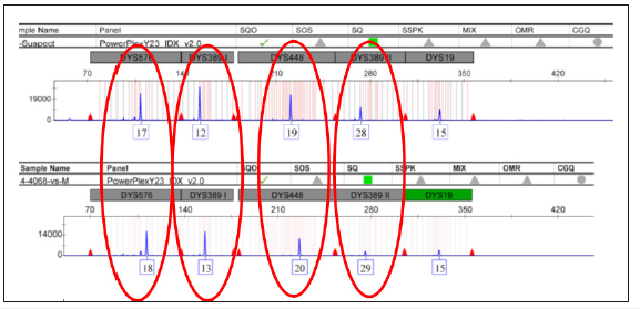
Figure 1:Comparison of YSTR DNA profile of panel no. 1 between victim and suspect.

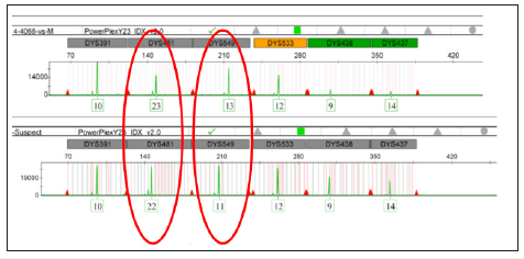
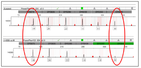
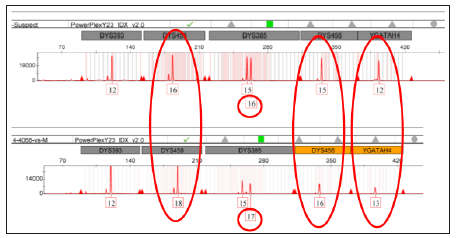
In the first phase of routine examination, Y STR kit was applied on the DNA of male fraction obtained by differential extraction method from the source of victim. We got identical male STR DNA profile from underwear, vaginal swab and vaginal slide that were indicating the presence of seminal stain on the source of victim. This result was also sustaining the outcome of biological examination in which the presence of spermatozoa was confirmed. These male DNA profiles were matched with the male STR profile from referral blood sample of suspect and we obtained astonishingly result (Figure 1-4). The alleles of the STR DNA profile of suspect’s source were not found similar with the alleles of male STR DNA profiles detected on the source of victim. This result was indicating that male stain found on the exhibits of victim was not source of suspect. We also performed autosomal STRs analysis and got same finding.
Figure 2:Comparison of YSTR DNA profile of panel no. 2 between victim and suspect.

Figure 3:Comparison of YSTR DNA profile of panel no. 3 between victim and suspect.

Figure 4:Comparison of YSTR DNA profile of panel no. 4 between victim and suspect.

Discussion
Sexual violence is a prevalent crime in today’s society needs some efforts to prevent it [10]. Enforcement of authoritarian commandment, fast track court for rapid decision and rigorous imprisonment or death penalty is the attempt that have implemented [11]. Every court trial in sexual assault case required vigilant investigation, examination and assessment for confirmation of suspect to be accused or innocent. Scientific evidence plays pivot role in making the mind of honorable judge to furnish their verdict (Human Right watch, 2010). In the last 50 years, microscopically detection of spermatozoa as a scientific tool has extensively been used in the examination of sexual assault cases [12]. It is often used to detect present of sperm cells on a victim or at a crime scene with a semen slide prepared from a suspect. If the sperm cell found on the source of victim along with on semen slide, not considered as strong evidence that sperm found on the source of victim came from the source of suspect. So, this outcome can’t say conclusive. With this finding if conviction is found to be unsafe then it treated as miscarriage of justice [13]. In present case study, suspect was found guilty in sexual assault case by the biology report mentioned that human spermatozoa were found on the source of victim. Suspect was penalized 10 years of rigorous imprisonment. When suspect filed petition for his DNA test then the same exhibits of the victim along with blood sample of suspect sent to laboratory. After examination we got result in which found that presence of male DNA profile on the source of victim not from the source of suspect.
On this report suspect was exonerate from the charge of sexual offence. Since 1992, the project name Innocence Project started with the using of DNA evidence. By this project approximately more than 365 prisoners have exonerated, in which also including twenty prisoners who were on death line and one of whom was only five days from putting to death (Innocence project). DNA profiling is a powerful forensic tool. It can be used to quickly eliminate a suspect, saving time in searches for perpetrators and it can provide compelling evidence to support a conviction and, most importantly, reduce the chances of a wrongful conviction [14,15].
Acknowledgment
I would like to give thanks to Mr. Dinesh Chandra Sagar, ADG, Technical Services, Home (Police) Department Madhya Pradesh & Dr. Harsh Sharma Director, State Forensic Science Laboratory, Sagar Madhya Pradesh for provides facility to complete this case work. I would like to give thanks to entire team of my DNA unit for their consistent support.
References
- Berger MA (2001) DNA and the criminal justice system: The technology of justice: John F Kennedy school of government: Lessons from DNA: Restriking the balance between finality and justice. Harvard University, USA.
- Jeffreys AJ, Wilson V, Thein SL (985) Individual specific fingerprint of human DNA. Nature 316: 76-79.
- Johnson P, Williams R (2004) Post-conviction DNA testing: The UK first exoneration case? Sci Justice 44(2): 77-82.
- Mishra T, Deepankar P (2018) Significance of DNA in Conviction of Rape Accused. International Journal of Advance Research and Development 3(3): 273-282.
- Gill P, Brenner C, Brinkmann B, Budowle B, Carracedo A, et al. (2001) DNA commission of the international society of forensic genetics: recommendations on forensic analysis using Y-chromosome STRs. Forensic Sci Int 124(1): 5-10.
- Wilson Wilde L (2005) DNA Profiling in criminal investigations in Freckleton and Selby, Expert Evidence. The Law Book Company 4: 80- 2053.
- Gerald LP (2018) Wrongful convictions and DNA exonerations: Understanding the role of forensic science. NIJ 279.
- Gill P, Jeffreys AJ, Werrett DJ (1985) Forensic application of DNA fingerprints. Nature 318: 577-579.
- Reid GA (1989) Molecular Cloning: A laboratory manual. In: Sambrook, Fritsch, Maniatis (Eds.), (2nd edn), Cold Spring Harbor Laboratory Press, USA, 3: E3-E4.
- Reid CT (2005) International review of modern sociology. JSTOR 31(2): 293–295.
- Kriti S (2004) Violence against Women and the Indian Law. In Savitri G (Ed.), Violence, Law and Women’s Rights in South Asia, Sage Publications, Thousand Oaks, California, USA.
- Allery JP, Telmon N, Mieusset R, Blanc A, Rougé D (2000) Cytological comparison of spermatozoa: Comparison of three staining methods. J Forensic Sci 46(2): 349-351.
- Naughton M (2006) Wrongful convictions and innocence projects in the UK: help, hope and education. Journal of Current Legal Issues.
- Sibille I, Duverneuil C, Grandmaison LG, Guerrouache K, Teissière F, et al. (2002) Y-STR DNA amplification as biological evidence in sexually assaulted female victims with no cytological detection of spermatozoa. Forensic Sci Int 125(2-3): 212-216.
- (2010) Dignity on trial: India’s need for sound standards for conducting and interpreting forensic examinations of rape survivors. Human Rights Watch.
© 2019 Praveesh Bhati. This is an open access article distributed under the terms of the Creative Commons Attribution License , which permits unrestricted use, distribution, and build upon your work non-commercially.
 a Creative Commons Attribution 4.0 International License. Based on a work at www.crimsonpublishers.com.
Best viewed in


.jpg)


 Editorial Board Registrations
 Submit your Article
 Refer a Friend
 Advertise With Us
.jpg)


.jpg)


.bmp)
.jpg)
.png)
.jpg)


.jpg)


.png)

.png)


.png)


