- Submissions

Full Text
COJ Nursing & Healthcare
The Conjoined Allies-Anastomosing Hemangioma
Anubha Bajaj*
Department of Histopathology, AB Diagnostics, India
*Corresponding author: Anubha Bajaj, Department of Histopathology, AB Diagnostics, New Delhi, India
Submission: April 25, 2023Published: May 05, 2023

ISSN: 2577-2007Volume8 Issue3
Opinion
Anastomosing haemangioma is a benign, vascular neoplasm which is reminiscent of and requires distinction from well differentiated angiosarcoma. Initially scripted within genitourinary tract, neoplasm may be delineated within diverse soft tissue sites and exhibits a predilection for para-spinal areas. Tumefaction is constituted of anastomosing cords of sinusoidal capillaries and vascular articulations layered by endothelial cells with minimal anisonucleosis along with an admixture of scattered hobnailed endothelial cells. Conservative surgical extermination of the neoplasm is curative. Anastomosing haemangioma is predominantly discerned within soft tissue, genitourinary tract, gastrointestinal tract or hepatic parenchyma. Grossly, neoplasm is well demarcated and non-encapsulated. Cut surface is hemorrhagic, spongy and appears mahogany brown [1,2].
Upon microscopy, neoplasm demonstrates a non-lobular architecture and is configured of anastomosing, proliferating capillaries and vascular articulations. Tumor architecture simulates splenic sinusoids and exhibits capillaries and vascular structures enmeshed within a framework of non-endothelial, supporting cellular component [1,2]. Layering endothelial cells display mild anisonucleosis and are intermixed with disseminated, hobnailed endothelial cells. Characteristically, fibrin thrombi are discerned. Mitotic activity is minimal to absent. Foci of extramedullary hematopoiesis and mature adipose tissue may be encountered in ~50% instances [1,2].
Upon microscopy, the well demarcated, non-encapsulated neoplasm is composed of loosely configured, lobular tumor architecture [3,4]. Low power magnification exemplifies alternating foci of cellular and pauci-cellular zones. Neoplasm is constituted of anastomosing, sinusoidal capillaries or vascular articulations. Vascular configurations are layered by hobnail endothelial cells. Layering endothelial cells may be focally flattened within specific zones [3,4]. Neoplastic cells appear devoid of cellular or nuclear atypia, multi-layering of endothelial cells, apoptotic bodies or mitotic figures. Mild nuclear enlargement may be observed [3,4]. Intervening stroma is scantily infiltrated by a population of lymphocytes. However, a component of infiltrating plasma cells or acute inflammatory cells are absent. Focal collagen deposition appears commingled with sinusoidal vascular articulations. Zones of sclerosis are encountered. Foci of vascular tumor invasion, tumor necrosis or infiltration into abutting adipose tissue, soft tissue or anatomic structures is absent [3,4]..
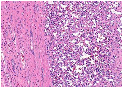
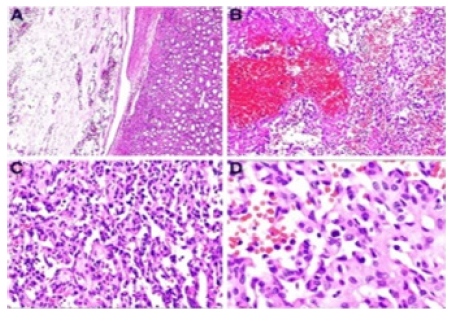
Anastomosing haemangioma is immune reactive to endothelial markers as CD31, CD34 and ERG (Figure 1). Tumor cells are diffusely immune reactive to CD31 and CD34. Stromal cells appear immune reactive to Smooth Muscle Actin (SMA) [5,6]. Anastomosing haemangioma is immune non-reactive to human herpes virus 8 (HHV8) [5,6] (Figure 2). Ki67 proliferation index is minimal and appears at ~1%. Anastomosing haemangioma necessitates segregation from neoplasms such as angiosarcoma, retiform hemangioendothelioma, hobnail haemangioma and splenic tissue configuring accessory spleen or focal splenosis [5,6]. Conservative management with simple surgical eradication of neoplasm is an optimal and recommended therapeutic strategy [5,6].
Figure 1:Anastomosing hemangioma delineating capillaries and vascular articulations lined by endothelial cells demonstrating mild anisonucleosis commingled with hobnail endothelial cells. Intervening stroma depicts scant, mature lymphocytes.

Figure 2:Anastomosing hemangioma exhibiting capillaries and vascular articulations lined by endothelial cells with mild anisonucleosis admixed with a smattering of hobnail endothelial cells. Anastomosing vascular configurations are intermingled with scanty population of small lymphocytes and foci of hemorrhage with red cell extravasation.

References
- McHenry A, Buza N (2023) Anastomosing hemangioma of the ovary with Leydig cell hyperplasia: A clinicopathologic study of 12 cases. Int J Gynecol Pathol 42(2): 167-175.
- Rogers T, Shah N, Mauro D, McGinty KA (2022) Anastomosing hemangioma of the liver: An unusual variant in abdominal MRI imaging. Radiol Case Rep 17(12): 4889-4892.
- Chang Chien YC, Beke L, Méhes G, Mokánszki A (2022) Anastomosing haemangioma: Report of three cases with molecular and immunohistochemical studies and comparison with well-differentiated angiosarcoma. Pathol Oncol Res 28: 1610498.
- Xue X, Song M, Xiao W, Chen F, Huang Q (2022) Imaging findings of retroperitoneal anastomosing hemangioma: A case report and literature review. BMC Urol 22(1): 77.
- Lappa E, Drakos E (2020) Anastomosing hemangioma: Short review of a benign mimicker of angiosarcoma. Arch Pathol Lab Med 144(2): 240–244.
- Cheon PM, Rebello R, Naqvi A, Popovic S, Bonert M (2018) Anastomosing hemangioma of the kidney: Radiologic and pathologic distinctions of a kidney cancer mimic. Curr Oncol 25: e220–e223.
© 2023 Anubha Bajaj. This is an open access article distributed under the terms of the Creative Commons Attribution License , which permits unrestricted use, distribution, and build upon your work non-commercially.
 a Creative Commons Attribution 4.0 International License. Based on a work at www.crimsonpublishers.com.
Best viewed in


.jpg)


 Editorial Board Registrations
 Submit your Article
 Refer a Friend
 Advertise With Us
.jpg)


.jpg)


.bmp)
.jpg)
.png)
.jpg)


.jpg)


.png)

.png)


.png)


