- Submissions

Full Text
COJ Nursing & Healthcare
Report and Review of a Case of Fusarium Infection from KUT in The Middle of IRAQ
Wifaq Mahmood Ali Al Wattar*
Unit of clinical and communicable diseases research, Iraq
*Corresponding author: Wifaq Mahmood Ali Al Wattar, Unit of clinical and communicable diseases research, Iraq
Submission: December 14, 2019;Published: February 20, 2020

ISSN: 2577-2007Volume6 Issue1
Opinion
Fusarium species is a ubiquitous fungus that causes opportunistic infections, those isolates are universally found in the environment and cause infection in both humans and plants [1-5]. In humans, infection starts with the inhalation of Fusarium conidia or by direct contact with Fusarium conidia. Subsequently, conidia germinate and form filaments that invade the surrounding tissue when a suitable environment is offered, clinical presentation of fusariosis depends on the host’s immune status. Invasive infections, such as sinusitis, pneumonia, deep cutaneous infections are the commonest in immunocompramized cases, On the other hand, immunocompetent patients present more frequently with superficial infections, such as keratitis and onychomycosis [6-8].
38 years old female teacher living at AL-KUT city, Wasset ,Iraq presented to medical city at November 2019, complaining of recurrent attacks of productive cough, with yellowish thick sputum with foul smell, she had low grade fever, nasal stuffiness with repeated attacks of shortness of breath relieved by broncho- dilator inhaler and repeated courses of steroids therapy, she lived in a rural area and she tend to raise chicken in her house back yard, her condition worsen each winter.
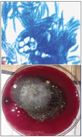
She came seeking medical advice in our hospital, her chest X-ray was normal with sight increase in bronchial marking, sputum sample was taken by medical mycology laboratory to be cultured on SDA media, blood agar and Brain heart agar. After two days of incubation multiple colonies of dark gray fluffy molds with elevated edges, wet mount was prepared to show banana shaped spores as shown in Figure 1. This is the first case to be diagnosed in our lab for 8 years of data collection with in 2500 cases.
Figure 1:

References
- Dignani MC, Anaissie E (2004) Human fusariosis. Clin Microbiol Infect 10(1): 67-75.
- Guilhermetti E, Takahachi G, Shinobu CS, Svidzinski TI (2007) Fusarium spp. as agents of onychomycosis in immunocompetent hosts. Int J Dermatol 46(8): 822-826.
- Guimera MNF, Garcia BM, Noda CA, Gonzalez RS, Montelongo RG (2004) Cutaneous infection by Fusarium: Successful treatment with oral voriconazole. Br J Dermatol 150(4): 777-778.
- Hsiue HC, Ruan SY, Kuo YL, Huang YT, Hsueh PR (2010) Invasive infections caused by non-Aspergillus moulds identified by sequencing analysis at a tertiary care hospital in Taiwan, 2000-2008. Clin Microbiol Infect 16(8): 1204-1206.
- Nucci M, Anaissie E (2006) Emerging fungi. Infect Dis Clin North Am 20(3): 563-579.
- Nucci M, Anaissie E (2007) Fusarium infections in immunocompromised patients. Clin Microbiol Rev 20(4): 695-704.
- Nucci M, Marr KA (2005) Emerging fungal diseases. Clin Infect Dis 41(4): 521-526.
- Chang DC, Grant GB, O Donnell K, Wannemuehler KA, Rao CY, et al. (2006) Multistate outbreak of Fusarium keratitis associated with use of a contact lens solution. JAMA 296(8): 953-963.
© 2020 Wifaq Mahmood Ali Al Wattar. This is an open access article distributed under the terms of the Creative Commons Attribution License , which permits unrestricted use, distribution, and build upon your work non-commercially.
 a Creative Commons Attribution 4.0 International License. Based on a work at www.crimsonpublishers.com.
Best viewed in


.jpg)


 Editorial Board Registrations
 Submit your Article
 Refer a Friend
 Advertise With Us
.jpg)


.jpg)


.bmp)
.jpg)
.png)
.jpg)


.jpg)


.png)

.png)


.png)


