- Submissions

Full Text
COJ Nursing & Healthcare
Cervical Stenosis - An Unusual Clinical Presentation
Kathpalia SK1*, Madhulima Saha2 and Namrita Sandhu2
1Andaman Nicobar Islands Institute of Medical Sciences, India
2Army College of Medical Sciences New Delhi, India
*Corresponding author: Kathpalia SK, Professor and HOD (Obstetrics and Gynecology), Andaman Nicobar Islands Institute of Medical Sciences, Allahabad, India, Email: kathpaliasukesh@gmail.com
Submission: February 05, 2018;Published: March 19, 2018

ISSN: 2577-2007Volume1 Issue5
Abstract
Outflow tract obstruction of female genital tract can lead to varied presentations depending on many factors. Most of the obstructions are congenital and usually occur in the lower part. A rare case of obstruction of cervical opening is reported. 37 years old lady with two previous cesarean deliveries reported with complaints of lower abdominal pain along with difficulty in passing urine, she was detected to have a lower abdominal lump. Imaging studies suggested retention cyst. Examination under anesthesia showed obliterated external cervical opening. Dilatation resulted in extrusion of large quantity of chocolate material. She made good postoperative recovery but the cause of cervical stenosis was not clear.
Keywords: Cervical stenosis; Hematometra; Urinary retention
Introduction
Outflow tract obstruction of female genital tract can lead to varied presentations depending on the site of obstruction, cause of obstruction and the age at the time of manifestation. Menstrual blood gets collected proximal to the obstruction and this collection can result in amenorrhea (cryptomenorrhoea), abdominal pain and pressure symptoms on lower urinary tract anteriorly and rectum posteriorly. Most of the obstructions are congenital in nature as imperforate hymen but some may be acquired also like after cervical electrocoagulation. We present an unusual case of acquired cervical stenosis which was a diagnostic dilemma.
Case Report
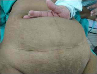
37 yrs old lady with two previous deliveries reported with complaints of pain in lower abdominal area along with difficulty in passing urine of 4 to 5 months duration. Pain was constant but got worse while passing urine, there was increased frequency of micturition and sense of incomplete evacuation but there was no burning while passing urine. She had two living children; both delivered by CS, last CS was performed 8 yrs back. The couple was using male condom as a method of contraception. Her menstrual cycles were absent for the last six to seven months but occasionally she had complained of altered blood stained vaginal discharge. General and systemic examination was essentially normal. Abdominal examination revealed a lump corresponding to 22 weeks size of pregnancy (Figure 1). The mass was firm to cystic in consistency. On internal examination the cervix and uterus could not be felt but a large mass was felt in the vagina, continuous with abdominal mass; whose mobility was restricted.
Figure 1: Large lower abdominal mass arising from pelvis.

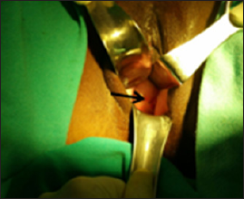
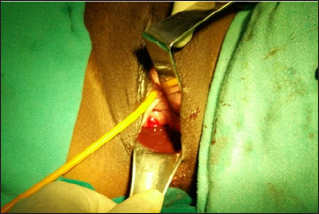
The mass appeared to be arising from anterior vaginal wall. A provisional diagnosis of anterior vaginal wall cyst was made and imaging studies performed. The ultrasound was suggestive of retention cyst (Figure 2). CT scan too pointed towards retention vaginal cyst (Figure 3). Since the diagnosis was not clear; the case was taken up for detailed examination under anesthesia, the mass was occupying the whole of vagina hence one could not go beyond the mass to locate the cervix, detailed per speculum examination under anesthesia revealed a small discolored spot (Figure 4) which when probed exuded tarry material (Figure 5). A diagnosis of cervical stenosis with retained blood was made and cervical opening was dilated to allow the collected blood to drain out. Appx 700 ml (Figure 6) of dark chocolate material was removed and cervix attained almost normal appearance and now the uterus could be palpated distinctly which appeared of normal size and shape. The lump abdomen had disappeared.
Figure 2: USG suggestive of retention cyst.

Figure 3: CT scan showing retention vaginal cyst.

Figure 4: Discolored cervical opening being shown by arrow.

Figure 5: Chocolate material being drained.

Figure 6: 700 ml of chocolate material drained.

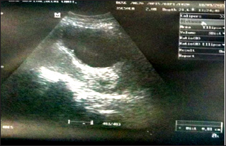
Figure 7: USG showing normal uterus and cervix 48 hrs after drainage

Ultrasound repeated 48 hrs later showed normal uterus with bulky cervix (Figure 7) indicating that the collection of blood was in cervical canal. Postoperatively the patient had uneventful recovery and her symptoms had disappeared; she was pain free and was able to pass urine normally. She was examined after her next menstrual cycle, per speculum examination was normal, and endocervical curettage did not show any evidence of malignancy. She has been followed up for six months, has been menstruating regularly and there is no recurrence.
Discussion
Development of female genital tract is highly complex and can result in many malformations of either fusion or recanalisation defects. These abnormalities are rare; [1] commonest obstruction of female genital tract is imperforate hymen, which results in accumulation of menstrual blood in the vagina resulting in hematocolpos. Diagnosis is usually made in childhood or at menarche with onset of cyclical pain and primary amenorrhoea [2]. Uncommon presentations include urinary retention [3], recurrent urinary tract infection or backache [4]. Hematocolpos can also occur in elderly women following vaginal occlusion secondary to radiotherapy or labial synechiae as a result of inflammatory conditions [5]. Cervical stenosis is a rare condition and can result from many causes like congenital cervical stenosis, chronic infection, surgical trauma like cone biopsy [6,7] cryotherapy or due to a tumor mass like a polyp or cervical cancer, post radiation or cervical endometriosis.
One rare cause of cervical stenosis has been described as CS [8], our patient too had undergone two CSs but her symptoms were not antedating surgery. The cause of such severe stenosis of cervix in our case was not clear. One of the differential diagnoses was cervical endometriosis but that was unlikely as there were no discolored nodules on the cervix and symptoms of endometriosis were absent. Cervical stenosis after cone biopsy is a recognized but rare complication [9]. Uterine cervical os [10], may appear normal, invisible, or may show a dimple, or a blue bulging membrane, in our case discolored opening was visible. Most of the cases can be diagnosed by ultrasonography; CT and MRI are rarely required [11]. Treatment is simple dilatation but at times recurrence [12] occurs for which different modalities have been used like catheter, stent or intrauterine device [13].
References
- Tran AT, Arensman RM, Falterman KW (1987) Diagnosis and management of hydrohematometrocolpos syndromes. Am J Dis Child 141(6): 632-634.
- McIvor RA (1990) Hematocolpos once in a lifetime cause of recurrent abdominal pain. Arch Emerg Med 7(1): 51-52.
- Hall DJ (1999) An unusual case of urinary retention due to imperforate hymen. J Accid Emerg Med 16(3): 232-323.
- Buick RG, Chowdhary SK (1999) Backache: a rare diagnosis and unusual complication. Pediatr Surg Int 15(8): 586-587.
- Speas CK, Gallup DC, Gallup DG (1998) Hematocolpos in elderly women. South Med J 86(7): 815-818.
- Xiaoqi Li, Jin Li, Xiaohua Wu (2015) Incidence, risk factors and treatment of cervical stenosis after radical trachelectomy: A systematic review. Eur J Cancer 51(13): 1751-1759.
- Borgatta L, Sayegh R, Betstadt SJ, Stubblefield PG (2009) Cervical obstruction complicating second-trimester abortion: treatment with misoprostol. Obstat Gynecol 113(2 Pt 2): 548-550.
- Kharat A, Kumar S (2014) Cervical Stenosis: a Rare Complication of Cesarean Section. Indian Journal of Clinical Practice 24(11): 1046-1047.
- Giannacopoulos K, Troukis E, Constandinou P, Rozis I, Kokonakis C, et al. (1998) Hematometra and extended vaginal haematoma after laser conization. A case report. Eur J Gynaecol Oncol 19(6): 569-570.
- Rosenberg ER, Trought WS (1981) The ultrasonographic evaluation of large cystic pelvic massed. Am J Obstet Gynecol 139: 579-586.
- Reuter KL, Young SB, Daly B (1994) Hematometra complicating conization with radiologic correlation. A case report. J Reprod Med 39(5): 408-410.
- Mathew M, Mohan AK (2008) Recurrent Cervical Stenosis - a Troublesome Clinical Entity. Oman Med J 23(3): 195-196.
- Caceres A, Jackson T (2012) Recurrent Hematometra Secondary to Cervical Stenosis Treated with Placement of an Intrauterine Device. The Journal of Minimally Invasive Gynecology 19(6): S160-S161.
© 2018 Kathpalia SK, et al. This is an open access article distributed under the terms of the Creative Commons Attribution License , which permits unrestricted use, distribution, and build upon your work non-commercially.
 a Creative Commons Attribution 4.0 International License. Based on a work at www.crimsonpublishers.com.
Best viewed in


.jpg)


 Editorial Board Registrations
 Submit your Article
 Refer a Friend
 Advertise With Us
.jpg)


.jpg)


.bmp)
.jpg)
.png)
.jpg)


.jpg)


.png)

.png)


.png)


