- Submissions

Full Text
COJ Biomedical Science & Research
Recent Advances in Quinoline Based Fluorescent Probes for Bio-imaging Applications
Jagriti Singh1* and Shachi Mishra2*
1School of Health Science and Technology, Indian Institute of Technology, Guwahati, Assam, India
2Assistant Professor, P. G. Department of Chemistry, Jai Prakash University, Chapra, Bihar, India
*Corresponding author: Jagriti Singh, School of Health Science and Technology, Indian Institute of Technology, Guwahati, India and Shachi Mishra, Assistant Professor, P. G. Department of Chemistry, Jai Prakash University, Chapra, 841302, Bihar, India
Submission: January 28, 2023; Published: February 22, 2023

Volume2 Issue3February , 2023
Abstract
Bioimaging has emerged as a prominent non-invasive technique for early diagnosis of several diseases especially cancer and neurodegenerative diseases. A lot of efforts have been devoted for the development of appropriate fluorescent probes which have great utility for experiments including cellular staining, tracking biomolecules of interest and, detection of specific bioanalytic in the cellular organisms. Quinoline-based scaffolds are among one of the widely explored moieties as molecular probes and chemosensory due to their interesting biological, photophysical and pharmacological properties. Herein, we have discussed some quinolone based fluorescent probes for bio-imaging applications.
Keywords:Quinoline; Fluorescent probes; Bio-imaging
Abbreviations:MPF: Multiphoton Fluorescent; 3PA: Three Photon Absorption; ICT: Intramolecular Charge Transfer; ESIPT: Excited State Intramolecular Proton Transfer; FRET: Forster Resonance Energy Transfer; PET: Photo-Induced Electron Transfer; CHEF: Chelation Enhanced Fluorescence; ROS: Reactive Oxygen Species
Introduction
Fluorescence imaging is a growing interest of field due to low cost, easy handling, high sensitivity, specificity, and target orientation [1,2]. It is an advanced non-invasive imaging technique that can detect, visualize, and characterize morphological and dynamic phenomena of biology at the molecular level in multi-dimensional fashions. Several fluorescent probes have slowly and steadily evolved and are now indispensable imaging tools in the field of molecular biology and medicine [3,4]. Among them quinoline-based fluorescent probes have been extensively explored due to their interesting biological, photophysical and pharmacological properties. In this mini review, we aim to summarize recent developments in quinoline-based fluorescent probes for bioimaging applications.
Quinoline-based scaffolds and their bioimaging application
Quinoline consists of benzene ring fused with a pyridine moiety, also known as 1-azanapthalene and benzo[b]pyridine [5]. It belongs to a privileged class of heterocyclic aromatic compound and exhibits a broad range of biological and pharmaceutical activities such as antibacterial, antioxidant, anticancer, anti-inflammatory, antimalarial, and antifungal [6-10]. In addition, it possess interesting fluorescence properties, which has been utilized in the development of various quinoline appended fluorescent molecules and has been exploited in the field of bio-imaging as molecular probes/chemo-sensors for monitoring the interactions with target molecules through the changes in fluorescence emission/intensity [11-19].
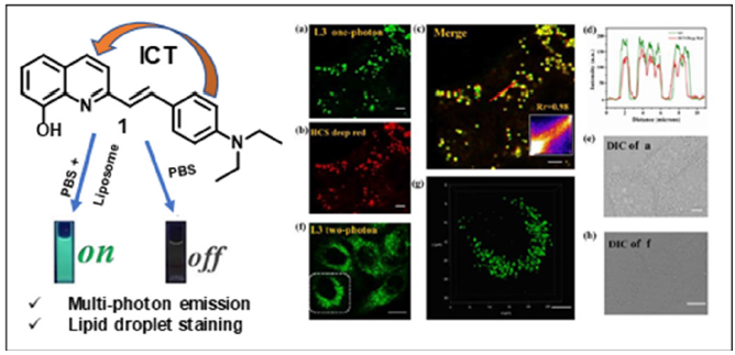
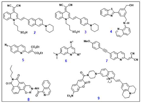
In 2020, Tian et al. [13] synthesized a series of quinoline based multiphoton fluorescent probe (MPF) for selective detection of lipid droplets in live cells. This MPF probe 1 exhibited ICT effect, deeper tissue penetration, lower autofluorescence and lower photo-toxicity (Figure 1). The three-photon absorption, 3PA cross sections of 1 was significantly enhanced when it binds to liposome and on the other hand due to large stokes Shift, it was highly selective to lipid droplets in living cells [13]. For detection and imaging of Aβ aggregates in Alzheimer’s disease, Yi et al. reported a water soluble two quinoline-malononitrile-based NIR fluorescent probes 2 and 3. These NIR probes were demonstrated for imaging of Aβ aggregates in in-vitro and in brain sections from triple transgenic mice of AD [14]. A turn-on quinoline and benzothiazole based chemo-sensor 4 was developed for the detection of Hg2+ through ESIPT mechanism. This chemo-sensor was successfully applied for the detection of Hg2+ in HeLa cells. Song et al. [16] reported quinoline containing two-input fluorescent probe (5, QME-N3) with two distinct reactive sites, the α, β-unsaturated carbonyl at 2-site and 6-azido group, which were specific acceptors of RSH and H2S respectively in live cells [16]. Chenoweth and his group rationally designed and synthesized a series of highly tunable quinoline-based small fluorescent molecule 6 and explored for various applications such as tunable photophysical properties and live-cell imaging followed by fluorescence response to intracellular pH [17]. During cellular diffusion-controlled processes, intracellular viscosity participates in the transportation and interaction of biomolecules and also abnormalities in viscosity produces many dysfunctions, such as Alzheimer’s disease and atherosclerosis [18] (Figure 2).
Figure 1:Structure of ICT based fluorescent probe 1 and Confocal images of live HeLa cells.

Figure 2:Structure of quinoline based fluorescent molecules for bio-imaging.

To visualize viscosity, Meng et al. [18] developed a twophoton fluorescent probe 7 which displayed off-on response with the increase of viscosity and also demonstrated imaging of intracellular viscosity in HeLa cells and zebrafish. In 2022, Xu et al. [19] discovered a reversible ‘‘turn-on” fluorescent probe 8 (NIQ) based on naphthalimide appended isoquinoline Schiff base for the detection of Al3+ in living cells. The sensing process involves inhibition of photo-induced electron transfer (PET) and the chelation-enhanced fluorescence (CHEF) process [20]. Sulfur dioxide (SO2) displayed important role in the regulation of insulin levels in the blood and lower blood pressure, but an elevated level also causes several diseases such as diarrhea, low blood pressure and cancer, neurological disease and cardiovascular disease. In order to detect endogenous and exogenous HSO3-/SO3 2- in live cells, recently Liu and his group synthesized near-infrared FRET fluorescent probe 9 which consisted of coumarin-quinolinejulolidine skeleton. This probe showed potential for differentiating normal cells from cancer cells and also targeting lysosomes [21].
Conclusion
This review discusses some recently developed quinolinebased fluorescent probes and highlights their potential use in cellular systems as imaging agents targeting liposomes, lysosomes, Aβ aggregates, intracellular pH and viscosity. Further, quinolinebased chemo-sensors for detection of toxic metal ions such as Hg2+ and Al3+ and reactive oxygen species (ROS) like RSH, H2S and HSO3 -/SO3 2- have also been designed and developed successfully. These probes showed advantages of a wide linear range, high quantum yields and low detection limit. Thus, these probes can help in early diagnosis of diseases like cardiovascular, cancer, atherosclerosis and neurodegenerative diseases, thereby forming the basis of most medical decision-making and limiting healthcare costs.
References
- Weissleder R, Mahmood U (2001) Molecular imaging. Radiology 219(2): 316-333.
- Ren M, Zhou K, He L, Lin W (2018) Mitochondria and lysosome-targetable fluorescent probes for HOCl: Recent advances and perspectives. Journal of Material Chemistry B 6(12): 1716-1733.
- Kobayashi H, Ogawa M, Alford R, Peter LC, Yasuteru U (2010) New strategies for fluorescent probe design in medical diagnostic imaging. Chemical Review 110(5): 2620-2640.
- Tamura T, Hamachi I (2019) Chemistry for covalent modification of endogenous/native proteins: From test tubes to complex biological systems. Journal of American Chemical Society 141(7): 2782-2799.
- Weyesa A, Mulugeta E (2020) Recent advances in the synthesis of biologically and pharmaceutically active quinoline and its analogues: A review. RSC Advance 10: 20784-20793.
- Vivekanand B, Raj KM, Mruthyunjayaswamy BHM (2015) Synthesis, characterization, antimicrobial, DNA-cleavage and antioxidant activities of 3-((5-chloro-2-phenyl-1H-indol-3-ylimino)methyl)quinoline-2(1H)-thione and its metal complexes. Journal of Molecular Structure 1079: 214-224.
- Desai NC, Kotadiya GM, Trivedi AR (2014) Studies on molecular properties prediction, antitubercular and antimicrobial activities of novel quinoline based pyrimidine motifs. Bioorganic & Medicinal Chemistry Letters 24(14): 3126-3130.
- EI Feky SA, Abd Samii ZKEl, Osman NA, Lashine J, Kamel MA, et al. (2015) Synthesis, molecular docking and anti-inflammatory screening of novel quinoline incorporated pyrazole derivatives using the pfitzinger reaction II. Bioorganic Chemistry 58: 104-116.
- Lyon MA, Lawrence S, William DJ, Jackson YA (1999) Synthesis and structure verification of an analogue of kuanoniamine A. Journal of the Chemical Society, Perkin Transactions 1 4: 437-442.
- Kundu B, Das SK, Chowdhuri SP, Pal S, Sarkar D, et al. (2019) Discovery and mechanistic study of tailor-made quinoline derivatives as topoisomerase 1 poison with potent anticancer activity. Journal of Medicinal Chemistry 62(7): 3428-3446.
- Lavis LD, Raines RT (2008) Bright ideas for chemical biology. ACS Chemical Biology 3(3): 142-155.
- Laras Y, Hugues V, Chandrasekaran Y, Blanchard DM, Francine CA, et al. (2012) Synthesis of quinoline dicarboxylic esters as biocompatible fluorescent tags. Journal of Organic Chemistry 77(18): 8294-8302.
- Zhang S, Yang Z, Li M, Zhang Q, Tian X, et al. (2020) A multi-photon fluorescent probe based on quinoline groups for the highly selective and sensitive detection of lipid droplets. Analyst 145: 7941-7945.
- Xu M, Li R, Li X, Lv G, Li S, et al. (2019) NIR fluorescent probes with good water-solubility for detection of amyloid beta aggregates in Alzheimer’s disease. Journal of Material Chemistry B 7(36): 5535-5540.
- Sahana S, Mishra G, Sri S, Bharadwaj PK (2015) A 2-(2′-hydroxyphenyl) benzothiazole (HBT)-quinoline conjugate: A highly specific fluorescent probe for Hg2+ based on ESIPT and its application in bioimaging. Dalton Transaction 44(46): 20139-20146.
- Dai CG, Liu XL, Du XJ, Zhang Y, Song QH (2016) Two-input fluorescent probe for thiols and hydrogen sulfide chemo-sensing and live cell imaging. ACS Sensor 1(17): 888.
- Jun JV, Petersson EJ, Chenoweth DM (2018) Rational design and facile synthesis of a highly tunable quinoline-based fluorescent small-molecule scaffold for live cell imaging. Journal of American Chemical Society 140(30): 9486-9493.
- Moriarty PM, Gibson CA (2003) LDL apheresis and its effect on viscosity. Cardiovascular reviews & report 24: 321-325.
- Ning P, Dong P, Geng Q, Bai L, Ding Y, et al. (2017) A two-photon fluorescent probe for viscosity imaging in vivo. Journal of Material Chemistry B 5(15): 2743-2749.
- Xu H, Zhang S, Gu Y, Lu H (2022) Phthalimide appended isquinoline fluorescent probe for specific detection of Al3+ ions and its application in living cell imaging. Spectrochimica Acta Part A: Molecular and Biomolecular Spectroscopy 265: 120364.
- Liu FT, Han WW, Ren H, Yang WJ, Wang RN, et al. (2023) A ratiometric lysosome-targeted fluorescent probe for imaging SO2 based on the coumarin-quinoline-julolidine molecular system. Dyes and Pigments 210: 111017.
© 2023 Jagriti Singh and Shachi Mishra. This is an open access article distributed under the terms of the Creative Commons Attribution License , which permits unrestricted use, distribution, and build upon your work non-commercially.
 a Creative Commons Attribution 4.0 International License. Based on a work at www.crimsonpublishers.com.
Best viewed in


.jpg)


 Editorial Board Registrations
 Submit your Article
 Refer a Friend
 Advertise With Us
.jpg)


.jpg)


.bmp)
.jpg)
.png)
.jpg)


.jpg)


.png)

.png)


.png)


