- Submissions

Full Text
Cohesive Journal of Microbiology & Infectious Disease
Histopathological Study of Colibacillosis Due to Escherichia Coli in Camels in Algeria
Rahmoun DE1, Lieshchova MA2* and Mylostyvyi RV2*
1University of Mohamed Cherif Messaâdia, Souk Ahras, Algeria
2Dnipro State Agrarian and Economic University, Dnipro, Ukraine
*Corresponding author: Lieshchova MA, Dnipro State Agrarian and Economic, University, Sergii Efremov, Ukraine Mylostyvyi RV, Dnipro State Agrarian and Economic, University, Sergii Efremov, Ukraine
Submission: January 27, 2023; Published: February 22, 2023

ISSN 2578-0190 Volume6 issues3
Abstract
Colibacillosis caused by Escherichia coli, is an acute infectious disease, mainly affecting young animals of all types. This disease is characterized by the appearance of abundant diarrhea, septicemia, severe dehydration, inflammation of the mucous membrane of the gastrointestinal tract and serous membranes. Our research is based on the evidence of the components of the intestine of camels affected by colibacillosis in the region of El-Oued in the south-east of Algeria. With the technique of silver nitrate impregnation, we were able to show fibrin deposits accompanied by macrophages, lymphocytes and plasma cells, with very variable rates.
Keywords: Chamelon; Colibacillosis; Escherichia coli; Fibrin
Introduction
The causative agent of infection can be localized in the intestines, in the urinary and biliary tract, lungs, and in some cases in the mesenteric lymph nodes. The causative agent of this disease are pathogenic strains of Escherichia coli. This pathogen belongs to the Enterobacteriaceae family. E. coli is a typical representative of the normal microflora of the gastrointestinal tract, that is, it is an obligate inhabitant that is normally found in the intestine but cannot cause the development of an infection only under certain conditions. This is facilitated by a decrease in the natural resistance of the body, a violation of the conditions of feeding and keeping animals, therefore, colibacillosis is called a factorial disease that manifests itself in the presence of pathogens, that is i.e., infectious agents, and predisposing factors, as well as the safety and subsequent productivity of the animals. The existence of an inflammatory tissue in situ, which includes cells that defend themselves against bacterial and viral aggressions, as cited by the authors Bourlioux et al. [1]. Newborn babies are exposed to various factors of an infectious and non-infectious nature that contribute to the onset of diseases, reduce the intensity of growth, productivity, and in most cases lead to death.
Materials and Methods
The research was conducted in the El-Oued region of southeastern Algeria. The object of the study was calves less than 30 days old, showed signs of diarrhea and dyspepsia. Histological studies of histological sections of the intestines of dead animals, stained with silver nitrate Sections frozen in the cryostat were impregnated with silver nitrate, according to the method of [2], which gave the ability to visualize all major components of the intestines simultaneously clearly, which due to the architectonics of the reticular fiber networks. The study of the histological preparations was carried out using Olympus CX-41 and Leica DM 1000 optical microscopes (eyepiece x 10, lenses x 10, x 40. The research results were statistically processed and presented using Statistic 12.0 (Stat Soft Inc., USA). The probability of a difference in values in different groups of gut fragments was assessed using Student’s t-test (P<0.05) after testing for normality of distribution and difference in general deviations.
Results and Discussion
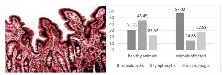
According to our research, the widespread diseases of young animals, accompanied by a violation of the digestive, secretory and absorptive function of the organs of the gastrointestinal tract, leading to diarrhea, are associated not only with altered feeding and maintenance of pregnant camels and young animals, but with the impact of certain infectious factors, and with exposure to the causative agent of dromedary diarrhea, colibacillosis. Colibacteriocins stands out among the main factorial diseases affecting the gastrointestinal tract of young dromedaries, which has been cited by the authors Hassan et al. [3] and having a massive character. The role of conditionally pathogenic microorganisms, mainly Escherichia, in the occurrence of this disease in young animals is steadily increasing. This disease causes important damages in the breeding [4]. The results of the anatomopathological study of the intestines of the dead animals, stained with silver nitrate, an increase in the quantity of fibrin was noticed by a deposition in clusters of reticulocytes with an accumulation of macrophage (Figure 1), large lymphocyte and plasmacyte. The analysis of the dynamics of the fibrin presence in relation to the Escherichia coli infection in camels, in both absolute figures and percentages, allows us to say that there was a decrease in the incidence of 57.02% of reticulocytes compared to the lymphocytes, which increased from a figure to 14.88%. The macrophages had a value of 27.58%.
Figure 1:Histological section small intestine of a young camel silver nitrate impregnation X 10 reticulocyte rate in the two groups of Animals (n=22).

The greatest fatality was noted in camels less than ten days old. Currently, among the main diseases of young farm animals under intensive breeding conditions, a special place is occupied by gastrointestinal diseases. The economic damage caused by ehrlichiosis consists of abortions and partial death of diseased females, death of young newborns, as well as the cost of important funds to carry out therapeutic, prophylactic, and anti-epizootic measures when eliminating the outbreaks of these diseases [4,5]. The formation of the fibrin caused by the inflammation of the intestinal mucosa is approved also on pig intestinal entero-bacteria according to the author [6].
Conclusion
In this research, the data allowed us to confirm that the disease of the intestine in the chameleon is fatal due to the presence of inflammation in situ with the presence of defense cells and fibrin deposit.
References
- Bourlioux P, Koletzko B, Guarner F, Braesco V (2003) The intestine and its microflora are partners for the protection of the host: report on the Danone Symposium. The American Journal of Clinical Nutrition 78(4): 675-683.
- Garvey W, Fathi A, Bigelow F, Carpenter B, Jimenez C (1989) A new method for demonstrating argyrophil cells of the pancreas and intestines. Stain Technology 64(2): 87-91.
- Hassan FA, Wernery U, Joseph M, Anouassi A, Mariena K (2019) Molecular identification of 20 Escherichia coli isolates from dead neonatal camel calves (Camelus dromedarius) in the United Arab Emirates. Journal of Camel Practice and Research 26(3): 259-260.
- Khalafalla AI, Hussein MF, Hussein MF (2021) Infectious diseases of dromedary camels: A Concise Guide, pp. 111-116.
- Khalafalla AI, Hussein MF (2021) Infectious diseases of dromedary camels. Cham, Springer International Publishing, Switzerland.
- Squire MM (2015) Clostridium difficile and idiopathic neonatal diarrhoea in Australian piglets (Doctoral dissertation, University of Western Australia.
© 2023, Lieshchova MA & Mylostyvyi RV. This is an open access article distributed under the terms of the Creative Commons Attribution License , which permits unrestricted use, distribution, and build upon your work non-commercially.
 a Creative Commons Attribution 4.0 International License. Based on a work at www.crimsonpublishers.com.
Best viewed in


.jpg)


 Editorial Board Registrations
 Submit your Article
 Refer a Friend
 Advertise With Us
.jpg)


.jpg)


.bmp)
.jpg)
.png)
.jpg)


.jpg)


.png)

.png)


.png)


