- Submissions

Full Text
Approaches in Poultry, Dairy & Veterinary Sciences
Angioarchitectural Study on the Intrahepatic Blood Supply of Geese (Anser Anser Domesticus) with Special Reference to its Biliary System
Amr F El Karmoty* and Ayman Tolba
Anatomy and Embryology department, Faculty of Veterinary Medicine, Egypt
*Corresponding author:Amr F El Karmoty, Anatomy and Embryology Department, Faculty of Veterinary Medicine, Egypt
Submission: May 25, 2019;Published: June 03, 2019
ISSN: 2576-9162 Volume6 Issue1
Abstract
The liver of geese bile ducts and its blood supply was demonstrated on ten specimens of adult healthy geese of different sex. The bile ducts were three; hepatoenteric, hepatocystic and cysticoenteric. The longer hepatoenteric duct was oriented by right, left and cranial hepatic dacutules distributed in the liver consistency. The cysticoenteric duct opens with the hepatoenteric duct in the duodenal papilla. The arterial supply of the liver goose is oriented by one right and two left hepatic arteries. The right hepatic artery branched into four craniodorsal, one caudoventral and anastomotic branch with the left hepatic artery. The left hepatic arteries arborized by one craniodorsal, two lateral and two caudoventral vessels. The intrahepatic venous drainage of the liver goose was established by hepatic portal and hepatic venous systems. The right hepatic portal vein distributed in the substance of its corresponding lobe where it anasomosed with the left hepatic portal vein via transverse part and gave off cranial, lateral and caudal tributaries. The left hepatic portal vein continued in the left lobe of liver detaching the common trunk of the cranial, lateral and caudal. The caudal vena cava was recieved blood via the middle hepatic vein and two main tributaries; right and left hepatic. Each vein it drained by craniodorsal, caudodorsal and ventral branches.
keywordsAngioarchitecture; Intrahepatic; Biliary; Geese
Introduction
Geese are from the lamellirostres because their bills have lamellae that fit with the bristles of the lateral margins of the tongue to sieve the food from the water [1,2]. Geese are used in guarding of the houses and belong to the tribe Anserini of the family Anatidae. This tribe comprises the genera Anser Anser Domesticus (grey geese), Branta (black geese) and Chen (the white geese). More distantly related members of the Anatidae family are swans, most of which are larger than true geese [3,4]. Published the economic importance of liver geese for production of foie gras.
The aim of our study is providing an accurate, anatomical descriptive bases of the intrahepatic blood supply and its biliary system that considered as a base for nutritionist for production of foie gras, for pharmacologist for elimination of some drugs as [5] as well as for surgeon for any surgical interference as removal of hepatic tumor.
Materials and Methods
Ten geese of different sex and weights were used in this study. Before exsanguinations, the birds were anaesthetized by IM injection of 0.5 cc of 2% xylazine HCL (3mg/kg) and followed by injection of heparin (Cal Heparin, 5000 I.U.) in the wing vein to prevent blood clotting. Each specimen was exsanguinated through the common carotid arteries and the jagular veins then left to bleed for 5 minutes. The breast muscles and the sternum were carefully removed for exposing liver. The cadavers were then preserved in a suitable container with 10% formalin solution for 3-5 days. Two of specimens were dissected to identify the morphological pattern of bile ducts and eight samples were injected by gum milk latex colored with red Rottring ink for the arteries and blue Rottring ink for the veins by cannulation through aorta and caudal vena cava. The injected specimens are preserved in 10% formalin solution for duration ranged between 8-10 days. The specimens were dissected for exposing the arterial supply and the venous drainage of the liver. The specimens were photographed using Olympus digital camera SP-600UZ 12 mega pixel. The nomenclature used in this study was that given by the Nomina Anatomica Avium [6].
Result
The main biliary system of liver geese consists of three bile ducts; hepatoenteric, hepatocystic and cysticoenteric. The hepatoenteric duct (Figure 1-3) is the longest one and measured about 3.2cm. It orients by right (Figure 2 & 3), cranial (Figure 3) and left [have cranial (Figure 3) and caudal (Table 1) branches] hepatic ductules to form a common hepatic duct which passes obliquely caudoventrally between the gall bladder and its fossa where it opens with the cysticoenteric duct in the upper third of the ascending duodenum forming a large elevated duodenal papilla. The hepatocystic duct (Figure 1-3) is the shortest and measured about 0.3cm where it directs obliquely caudodorsally where it opens in the ventromedial wall of the gall bladder to evacuate the bile. The cysticoenteric duct (Figure 1-4) originates from the dorsolateral wall of the gall bladder and passes caudoventral oblique to open with the hepatoenteric duct in the current papilla.
Figure 1:A photograph showing bile ducts of liver goose.
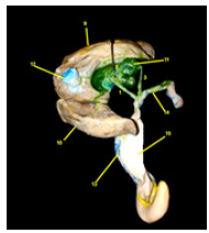
Figure 2:A photograph showing biliary system of liver goose.
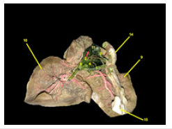
Figure 3:A photograph showing biliary system of liver goose.
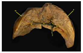
Figure 4:A photograph showing branches of celiac artery of goose.
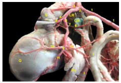
Figure 5:A photograph showing branches of celiac artery of goose.
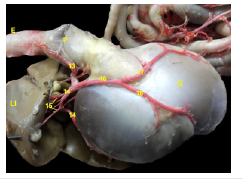
Figure 6:A photograph showing intrahepatic arterial supply of liver goose.

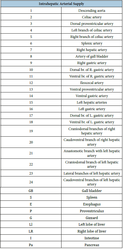
Table 1:

The arterial supply of the liver goose is oriented by one right and two left hepatic arteries. The right hepatic artery (Figure 4) is a long single branch which emanates from the right lateral wall of the right branch of the celiac artery and terminates in the porta hepatis of the right lobe of the liver. During its course it detaches artery of the gall bladder. The current artery is supplying the hepatic tissue of the corresponding lobe where it ramified into; four craniodorsal, one caudoventral and anastomotic branch with the left hepatic artery.
The first four craniodorsal branches directs obliquely in highly tourous manner to supply the cranial third of the right lobe of the liver. The caudoventral one is a long slender vessel measures about 4.9cm. It passes caudally to supply caudal two thirds of concurrent lobe. Along its course it gives off six collateral branches. The left hepatic arteries are two small vessels that arise from the dorsal wall of the ventral gastric artery and direct cranioventrally towards the left lobe of the liver where it arborized through its consistency by three groups of fine branches; one craniodorsal, two lateral and two caudoventral vessels that supplying the corresponding cranial, middle and caudal parts of the left lobe of the liver respectively (Table 2).
Table 2:
The intrahepatic venous drainage of the liver goose is established by hepatic portal and hepatic venous systems where the right and the left hepatic portal veins enter the liver from its visceral surface then ramify to form their tributaries within the consistency of liver tissue followed by forming the tributaries of the right and left hepatic veins that drain into the caudal vena cava. The right hepatic portal vein is a large vessel which measures about 3cm in its length. It directs to the right porta hepatis at the ventral border of the gall bladder where it receives the splenic, the gastropancreaticoduodenal and the common mesenteric veins. The current vessel distributed in the substance of its corresponding lobe where it anasomosed with the left hepatic portal vein via transverse part; have two branches; cranial and ventral.
Figure 7:A photograph showing tributaries of right hepatic portal vein of liver goose.
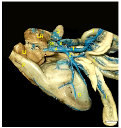
Figure 8:A photograph showing tributaries of left hepatic portal vein of liver goose.
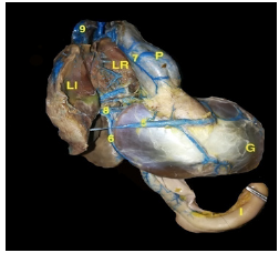
Figure 9:A photograph showing the intrahepatic architecture of the right and the left hepatic portal veins of liver goose.

Figure 10:A photograph showing the intrahepatic tributaries of the caudal vena cava of liver goose.

Table 3:
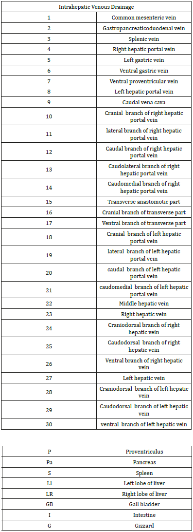
Along its course it gives off cranial, lateral and caudal tributaries where the latter one divides into the caudolateral and the caudomedial ones. The left hepatic portal vein is a small vessel which measures about 1.5. It passes to its porta after drained by left gastric, ventral gastric and ventral proventricular veins. It continues in the left lobe of liver detaching the common trunk of the cranial and lateral veins as well as a large caudal tributary that drained during its course by medial vessel. Finally, the caudal vena cava is received detoxified blood via the middle hepatic vein and two main tributaries; right and left hepatic. Each vein it drained by craniodorsal, caudodorsal and ventral branches that collect blood from all directions of their corresponding liver tissue.
Discussion
Our study revealed that the main biliary system of liver geese consists of three bile ducts; hepatoenteric, hepatocystic and cysticoenteric. The hepatoenteric duct is the longest one and measured about 3.2cm. It orients by three hepatic ductules; right, cranial and left which have cranial and caudal branches. The corresponding ductlues meet together forming a common hepatic duct which passes obliquely caudoventrally between the gall bladder and its fossa where it opens with the cysticoenteric duct in the upper third of the ascending duodenum forming a large elevated duodenal papilla. The hepatocystic duct is the shortest and measured about 0.3cm where it directs obliquely caudodorsally where it opens in the ventromedial wall of the gall bladder to evacuate the bile. The cysticoenteric duct originates from the dorsolateral wall of the gall bladder and passes caudoventral oblique to open with the hepatoenteric duct in the current papilla.
These results were simulated to that reported [2,7-11] asserted that the right and the left hepatic ducts connected on the visceral surface of the right lobe to form the common hepatoenteric duct which opened in the distal part of the descending duodenum via a small papilla. In owl, jackdaw and darter there was another right hepatoenteric duct which emanates from the right lobe of the liver and opened in the commencement of the descending duodenum. The gall bladder adhered to the common hepatic duct by a hepatocystic duct. A cysticoenteric duct derived from the gall bladder and opened in the distal part of the descending duodenum. In dove and sparrow a hepatoenteric duct arose from the common hepatic duct and meet to the ascending duodenum. However, [8,9] observed that there were only two hepatoenteric ducts ; the right duct emptied into the ascending limb and the largest left duct into the descending limb of the duodenum.
Moreover, [10] defined that there was another right hepatoenteric duct opened in the proximal part of the descending duodenum. The gall bladder attached to the right hepatic duct by the hepatocystic duct where from the gall bladder cysticoenteric duct arose and opened in the distal part of the ascending duodenum and in pigeon the right hepatoenteric duct opened directly into the ascending duodenum. While [12] said that there was no gallbladder and have a large hepatoenteric duct arose from the porta hepatis which opened into a papilla in the descending limb of the duodenum. Concerning the internal ductules of hepatoenteric duct , our study was differ from that stated by [13] where recorded that the common hepatoenteric duct was consisted of the right and left hepatic ducts that were divided into two parts; ventral and dorsal ductules.
Concerning the origin of the right hepatic artery was a long single branch which emanated from the right lateral wall of the right branch of the celiac artery and terminated in the porta hepatis of the right lobe of the liver. These results similar to that stated by [5-7,14-25] reported that both the right and left hepatic arteries are given off from the left branch of the celiac artery and terminated in the right and left lobes of the liver, respectively. However, [26-28] observed that the main branches of the celiac artery are superior and inferior gastric arteries that supplying the left and right hepatic arteries, respectively, to the liver lobes.
In our study recorded that the right hepatic artery was supplying the hepatic tissue of the corresponding lobe where it ramified into; four craniodorsal, one caudoventral and anastomotic branch with the left hepatic artery. The first four craniodorsal branches directed obliquely in highly tourous manner to supply the cranial third of the right lobe of the liver. The caudoventral one was a long slender vessel measured about 4.9cm. It passed caudally to supply caudal two thirds of concurrent lobe. Along its course it gave off six collateral branches. While [5] showed that the right hepatic artery inside the right hepatic lobe was as unique pattern of distribution where it was arborized into dorsal and ventral sets of arterioles that supplied the right lobe.
In the examined liver reported that the left hepatic arteries were two small vessels that arose from the dorsal wall of the ventral gastric artery and directed cranioventrally towards the left lobe of the liver. These statements were similar to that declared by [7,15,18,19,21,29-31] stated that the ventral gastric artery gives off the left hepatic artery, distributing to the left lobe of the liver. Added that the latter artery was a single vessel in ducks. These results which were in contrast to the statements that revealed by [5,20,25] where they described that the left hepatic artery emanated directly from the left branch of the celiac artery to the left lobe of the liver, while [26] said that the current branch detached directly from the celiac artery.
Regarding the distribution of the left hepatic artery was arborized through its consistency by three groups of fine branches; one craniodorsal, two lateral and two caudoventral vessels that supplying the corresponding cranial, middle and caudal parts of the left lobe of the liver, respectively. On the other hand [5] showed that a specific pattern of ramification where it arborized in a dorsoventral fashion; two dorsal, one ventral and two fine branches on the same plane inside the hepatic substance.
In the present study the hepatic portal venous system is oriented by right and left hepatic veins these results come in agreement with [7,27,32-34] in ducks and geese declared that the current venous system was constructed via two left and one right hepatic portal veins.
Regarding to the right hepatic portal vein was a large vessel which measured about 3cm in its length. It directed to the right porta hepatis at the ventral border of the gall bladder where it received the splenic, the gastropancreaticoduodenal and the common mesenteric veins. These results were simulated with that established by [7,33,35] added that there was the right proventricular vein which drained into the right portal vein. However [5] reported that the corresponding vessel was received by proventriculosplenic, gastropancreaticoduodenal and common mesenteric veins.
Concerning the distribution of the right hepatic vein was anasomosed with the left hepatic portal vein via transverse part; have two branches; cranial and ventral. Along its course it gave off cranial, lateral and caudal tributaries where the latter one divides into the caudolateral and the caudomedial ones. These statements might to be attributed to that noted by [32] although who reported that the transverse part gave cranial and caudal 1-6 branches. The latter statement was similar to that recorded by [34]. However the latter author showed that the right hepatic portal vein emerged the cranial and caudal branches only. On the other hand [5] described that the branches of the corresponding vein were dorsal, lateral and ventral ones.
In the current study the left hepatic portal vein was drained by ventral gastric, left gastric and ventral proventricular veins. These results were similar to the conducted by [5,7]. While [33] concluded that the left portal hepatic vein was formed by 2 left gastric, ventral gastric, the pyloric and the caudal proventricular veins.
The results under discussion achieved that the left hepatic portal vein was detached the common trunk of the cranial and lateral veins as well as a large caudal tributary that drained during its course by medial vessel. While [32] designated that the left hepatic portal vein gave cranial, lateral and medial branches. On the other hand [5] in turkey had the opinion that the corresponding branch emerged dorsal, lateral and ventral ones. However [34] stated that the current vein was received cranial, caudal and medial branches but in duck and goose was drained by cranial, caudal branches only.
In viewing of the present study, the caudal vena cava was received detoxified blood via the middle hepatic vein and two main tributaries; right and left hepatic. Each vein it drained by craniodorsal, caudodorsal and ventral branches that collect blood from all directions of their corresponding liver tissue. These results were simulated to that mentioned by [6,35].
Conclusion
The liver of geese bile ducts were three; hepatoenteric, hepatocystic and cysticoenteric. The longer hepatoenteric duct was oriented by right, left and cranial hepatic dacutules distributed in the liver consistency. The arterial supply of the liver goose was oriented by one right and two left hepatic arteries. The intrahepatic venous drainage of the liver goose was established by hepatic portal and hepatic venous systems. These anatomical data was a base for nutritionist for production of foie gras, for pharmacologist for elimination of some drugs and for surgeon for any surgical interference.
References
- Mally BO (2005) Clinical anatomy and physiology of exotic species (structure and function of mammals, birds, reptiles and amphibians. El Sevier Saunders; Edinburg London New York Oxford Phiadelphia Tt Louis Sydney Toronto, Canada.
- Mc Lelland J (1990) A colour atlas of avian anatomy. Philadelphia WB, London, Toronto, Montreal, Sydney, Tokyo, Japan.
- Barnes GG, Vernon G, Thomas V (1987) Digestive organ morphology, diet and guild structure of North American Anatidae. Canadian Journal of Zoology 65(7): 1812-1817.
- Guy G, Fortun LL, Benard G, Fernandez X (2013) Natural induction of spontaneous liver steatosis in graylag landaise geese (Anser anser). Journal of animal science 91(1): 455-464.
- EL Gammal SMM (2016) Anatomical studies on the blood vessels of the gastrointestinal tract of the Turkey, with Special Reference to the Hepatic Portal System and Biliary Elimination of some Drugs.
- Baumel JJ, King SA, Breasile JE, Evans HE, Berge JCV (1993) Nomina Anatomica Avium. Published by the Nuttall Ornithological Club. No: 23, Cambridge, Massachusetts, USA.
- El Karmoty FA (2014) Macromorphological study on the digestive system of geese and ducks with special reference to its blood supply. Cario University, Egypt.
- Dyce KM, Sack WO, Wensing CJG (2010) Textbook of veterinary anatomy (4th edn), ElSevier Saunders. pp. 1-864.
- El Shafey AFAM, El-shaieb MOH, Metwally MAM (2006) Some comparative anatomical studies on the stomach, intestine and liver in ducks, chicken and pigeon. PhD Thesis, Fac Vet Med, Benha University, Egypt.
- Ibrahim IA, Abdalla KEH, Mansour AA, Taha A (1992) Topography and morphology of the liver and biliary duct system in fowl, duck, pigeon, quail, heron and kestrel. Assuit Vet Med J 27(53): 12-32.
- Hassouna EMA, Zayed AE (2000) Some morphological and morphometerical studies on the liver and biliary duct system in goose, turkey, dove, sparrow, jackdaw, hoopoe, owl and darter. Assuit Vet Med J 44(88):
- Stornelli MR, Ricciardi MP, Giannessi E, Coli A (2006) Morphological and histological study of the ostrich (Struthio Camelus L.) liver and biliary system. Italian Journal of Anat Embryol 111(1): 1-7.
- Al-Sadi S, Al-Hasso BS (2014) Anatomical and radio graphic investigations of the gall bladder and biliary duct in geese (Anser banikaval) and ducks (Anas gallopavo). Iraqi Journal of Veterinary Sciences 28(2): Ar133- Ar141.
- Farag FMM, Tolba AR, Daghash SM (2013) The arterial supply of the intestinal tract of the domestic turkey fowl (meleagris gallopavo). Journal Vet Anat 6(1): 1-16.
- Ragab SA, Farag FMM, Tolba AR, Saleh AA, El-Karmoty AF (2013) Anatomical Study on the Celiac Artery in the Domestic Goose (Anser anser domesticus) with Special Reference to the Arterial Supply of the Stomach. Journal Vet Anat 1 6(2): 23-40.
- Aslan K, Takci I (1998) The arterial vascularisation of the organs (stomach, intestinum, splen, kidneys, testes and ovarium) in the abdominal region of the geese obtained from Kars surrounding (in Turkish). Kafkas University, Fac Vet Med J 4: 49-53.
- Malinovsky L, Visnanska M (1975) Branching of the celiac artery in some domestic birds, II. The domestic goose. Folia Morphologica (Prague) 23(2): 128-135.
- Kurtul I (2002) Comparative macroanatomical investigations on the pattern and branches of the aorta descendens among the rooster, drake, and pigeon (in Turkish). Fac Vet Med Ankara Univ, pp. 24-37.
- Dursun N (2002) Anatomy of Domestic Birds (in Turkish). Medisan Publishing, Ankara, pp. 140-141.
- Baumel JJ (1975) Aves Heart and Blood Vessels. In, Sisson and Grossman’s the Anatomy of the Domestic Animals. Getty R (Ed.), (5th edn), Saunders Company, Philadelphia 5: 1990-1991.
- Malinovsky L, Novotna M (1977) Branching of the coeliac artery in some domestic birds. III. A. Comparison of the pattern of the coeliac artery in three breeds of the domestic fowl (Gallus gallus f. domestica). Anat Anz 141(2): 136-146.
- McLeod WM, Trotter DM, Lumb JM (1964) Avian Anatomy. Burgeaa Publishing Company 82: 133-134.
- Malinovsky L, Visnanska M, Roubal P (1973) Branching of a. celiaca in some domestic birds. I. Domestic Duck (in Czech). Scripta Medica 46: 325-336.
- Malinovsky L (1965) Blood supply to stomachs and adjacent organs in Buzzard. Folia Morphologica (Praque) 13: 191-201.
- Haligur A, Duzler A (2010) Course and branch of the celiac artery in the red falcon (Buteo rufinus). Vet Med 55(2): 79-86.
- Aycan K, Duzler A (2000) The anatomy of celiac artery in the eagle owl (Bubo bubo) (in Turkish). Ankara University, Fac Vet Med J 47: 319-323.
- Nickel R, Schummer A, Seiferle E (1977) Anatomy of the Domestic Birds. Verlag Paul Parey, Berlin, Germany.
- Gadhoke JS, Lindsay RT, Desmond RK (1975) Comparative study of the major arterial branches of the descending aorta, and their supply to the abdominal viscera in the domestic turkey (Meleagris gallopavo). Anat Anz 138(5): 438-443.
- Kuru N (2010) Macroanatomic investigations on the course and distribution of the celiac artery in domestic fowl (Gallus gallus domesticus). Scientific Research and Essays 5(23): 3585-3591.
- Kuru N (1996) Macroanatomical investigation of course and branching of aorta in domestic chick and New Zealand rabbit (in Turkish). Faculty of Biology, Selcuk University. pp. 30-37.
- Franz V, Salomon V (1993) Lehrbuch der Geflügelanatomie. Gustav Fischer Verlag, Jena, Suttgart, Germany.
- Santos TC, Ferrari CC, Menconi A, Maia MO, Bombonato PP, et al. (2009) Veins from hepatic portal vein system in domestic geese. Veias do sistema porta-hepático em gansos domésticos. Pesquisa Veterinária Brasileira 29(4): 327-332.
- Pinto MRA, Ribeiro AACM, Souza WMD, Miglino MA, Machado MRF (1999) Estudo do sistema portal hepático no pato doméstico (Cairina moshata) {the study of the portal hepatic system in the domestic duck (Cairina moshata)} Brazilian Journal of Veterinary Research and Animal Science, Braz J Vet Res Anim Sci 36(4).
- Jiaji C (1997) The Hepatic Portal Venous System of Fowl. Chinese Journal of Veterinary Science 1997-2001.
- Nishida T, Paik YK, Yasuda M (1969) Blood vascular supply of the glandular stomach (Ventriculus glandularis) and the muscular stomach (Ventriculus muscularis). Jpn J Vet Sci 31: 51-70.
© 2019 Amr F El Karmoty. This is an open access article distributed under the terms of the Creative Commons Attribution License , which permits unrestricted use, distribution, and build upon your work non-commercially.
 a Creative Commons Attribution 4.0 International License. Based on a work at www.crimsonpublishers.com.
Best viewed in


.jpg)


 Editorial Board Registrations
 Submit your Article
 Refer a Friend
 Advertise With Us
.jpg)


.jpg)


.bmp)
.jpg)
.png)
.jpg)


.jpg)


.png)

.png)


.png)


