- Submissions

Full Text
Approaches in Poultry, Dairy & Veterinary Sciences
Cervical Nerve Plexus Block for Treatment of Acquired Spasmodic Cervical Torticollis (Scoliosis) in an Arabian Horse
Mohamed Shokry1* and Fawzy Elnady2
1 Department of Surgery, Anesthesiology & Radiology, Egypt
2 Department of Anatomy & Embryology, Egypt
*Corresponding author: Mohamed Shokry, Department of Surgery, Anesthesiology &Radiology, Faculty of Veterinary Medicine, Cairo University, Giza, Egypt
Submission: January 14, 2019;Published: January 31, 2019

ISSN: 2576-9162 Volume5 Issue5
Abstract
A case report of an Arabian horse presented with severe acquired cervical torticollis (scoliosis) and successfully treated after cervical nerve plexus block.
Introduction
In the horse, acquired cervical torticollis (scoliosis), in which the cervical vertebrae are twisted or crooked, appears to be rare with few reported cases [1,2]. A variety of reasons are reported for acquired cervical scoliosis. Musculo-skeletal causes have included fracture/subluxation of cervical vertebrae, unilateral cicatrical muscle contracture, unilateral muscle rupture, unilateral paralysis subsequent to cervical spinal nerve damage and nutritional dystrophic myo-degeneration [1]. This clinical report describes a technique for correction of an acquired cervical torticollis in a horse.
History
A 6-year-old male Arabian horse was presented to the surgery clinic of the faculty of Veterinary Medicine, Cairo University, with a history of 3 days duration of a deviated neck. The owner reported that the horse developed severe neck deviation following tying of the horse head with a short rope combined with unsuccessful attempts to release the hanged head with recumbence.
Clinical Findings
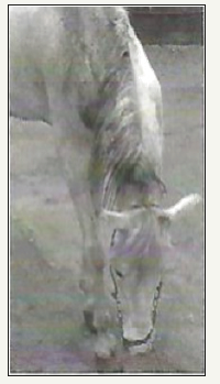
Physical examination revealed the presences of neck deep abrasion from the rope. There was severe torticollis of the neck to the left with inability to raise the head up. The concavity of the deviated neck occupied the right mid-neck side Figure 1. Neurological examination was normal. Radiography of the cervical vertebrae did not show any bony or articular lesions.
Figure 1:A 6-year-old Arabian horse with acquired cervical scoliosis.

Treatment
Dorsal cervical nerve plexus block
The horse was anesthetized and positioned in lateral recumbency on the left side. Aseptic preparation of the right concave side of the affected neck was performed. The wing of the atlas as well as the transverse process of the subsequent cervical vertebrae were palpated at the concave right side of the neck as far as caudally to the level of C6. With deep digital pressure, the intervertebral indentations between the cervical vertebrae were palpated and marked with a marker pen indicating the landmarks of the branches of the dorsal cervical nerve plexus from C3 to C6. A 12cm long and 21-gauge spinal needle was inserted through the skin at the first landmark (C3) in the intervertebral depression until it stuck the articular process of cervical vertebra. Little withdrawal of the needle allowed contact with the target nerve. Then 10ml of Mepivacaine HCl 2% and 40mg Triamcinolone Acetonide (Kinacort, Squibb, Egypt) suspension were injected and distributed in the vicinity of the nerve. The other landmarks of the subsequent dorsal cervical branches (C4, C5 & C6) were similarly treated Figures 2 & 3. Successful infiltration was indicated by full analgesia including the skin within 10 minutes associated with rise of skin temperature. By gentle massaging of the involved muscles of the neck, the twisted vertebrae assumed their normal position. A side stick between the head and neck collar was fixed while the head was tied to a high wall ring, eating from a high hay rack for 48 hours.
Figure 2:The exits of dorsal cervical nerve plexus (C3-C6) in relation to cervical vertebra.

Figure 3:The landmarks of injection sites.

Results
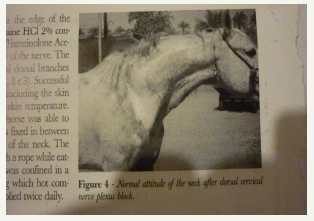
Fast improvement was achieved after 48 hours the neck of the horse assumed normal attitude Figure 4. The animal was discharged with instructions to continue box confinement and fixing of the head in high position, eating from a high rack, daily neck massaging with methyl sulfonyle methane (MSM ointment and oral phenylbutazone 4mg/kg bid for one extra week.
Figure 4:Normal attitude of the neck after treatment.

Discussion
From the case history, the presented report of acquired cervical torticollis after accidental trauma of the neck was related to severe spasmodic contraction of the lateral cervical muscles and the attached ligamentum nuchae without involvement of the vertebrae. Musculoskeletal and neurogenic caused were reported due to cervical nerve damage or dorsal grey column myelitis [1,2]. Recently, parasitic (verminous) sensory myelitis by strongylus tenuis was also implicated [2]. Early therapeutic measures should be considered in acquired cervical torticollis of non-neurogenic or skeletal origin since secondary focal articular changes in the articular processes were developed, varying from enlargement on the convex side to fibrosis with hemorrhage and granulation tissue formation on the concave or fractures of the articular processes of the vertebrae associated with abnormal loading and immobilization of the vertebral column [3]. The dorsal cervical plexus nerve block proved its efficiency and simplicity for treatment of acquired spasmodic torticollis of non-neurogenic or skeletal lesions in the horse. The dorsal cervical nerve plexus formed by connections of the dorsal branches of C3-C6 via communicating branches is the major nerve supply to the lateral aspect of the neck. The lateral branches of the dorsal branches of the cervical nerves from the main motor supply to the dorsolateral muscles of the neck while the medial branches supply the deep lateral muscles and the skin [4,5]. Injecting the local analgesic Mepivacaine HCl 2% releases the spaticity of the involved muscles of the neck and provides the requested analgesia for free mobility which helps the remodeling of the affected muscles the addition of potent steroidal anti-inflammatory Triamcinolone acetonide is very important to alleviate myositis and desmitis in the affected region.
References
- McKelvey WA, Owen RR (1979) Acquired torticollis in eleven horses. J Am Vet Med Assoc 175(3): 295-297.
- Biervliet VJ, Lahunta DA, Ennulat D, Oglesbee M, Summers B (2004) Acquired cervical scoliosis in six horses associated with grey column chronic myelitis. Equine Vet J 36(1): 86-92.
- Enneking WF, Harrington P (1969) Pathological changes in scoliosis. J Bone Joint Surg Am 51(1): 165-184.
- Getty R (1975) The anatomy of the Domestic Animals. (5th edn), WB Saunders Company, Philadelphia, London, Vol. 1.
- Sack WO (1991) Rooney’s Guide to the dissection of the horse. (6th edn), Ithaca, New York, USA.
© 2019 Mohamed Shokry. This is an open access article distributed under the terms of the Creative Commons Attribution License , which permits unrestricted use, distribution, and build upon your work non-commercially.
 a Creative Commons Attribution 4.0 International License. Based on a work at www.crimsonpublishers.com.
Best viewed in


.jpg)


 Editorial Board Registrations
 Submit your Article
 Refer a Friend
 Advertise With Us
.jpg)


.jpg)


.bmp)
.jpg)
.png)
.jpg)


.jpg)


.png)

.png)


.png)


