- Submissions

Full Text
Associative Journal of Health Sciences
An Interesting Presentation of Carcinoma Lung-Case Report
Dr A Nirmal kumar*, Dr A Arunkumar, Dr Javid Raja and Dr Rajkumar
Assistant Professor, Sri Manakula Vinayagar Medical College & Hospital, India
*Corresponding author: A Nirmal kumar, Assistant Professor, Department of CTVS, Sri Manakula Vinayagar medical college & hospital, Puducherry, India
Submission: July 01, 2021;Published: August 17, 2021

ISSN:2690-9707 Volume1 Issue4
Introduction
Sternal masses can be either primary or secondary with two thirds of masses resulting from metastasis [1]. Metastatic secondaries usually come from breast, lung, kidney and thyroid [2]. The skeleton is a common site of metastasis in bronchogenic carcinoma. However, sternal involvement is rare. We report a case of atypical presentation of lung carcinoma to increase the awareness of surgeons and physicians.
Case Report
Figure 1:

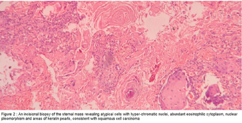
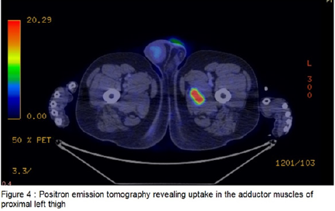
A 56-year-old male with a history of smoking 30 pack years presented with complaints of swelling over the upper sternum and pain over the swelling for the last three months. The pain was persistent, progressive and localized. There was no history of hemoptysis, fever, hoarseness of voice, or weight loss. However, loss of appetite was present. On inspection, a swelling of size 5.5×4cm was seen in the region of the manubrium. The sternal swelling was tender and firm in consistency. No cervical or generalized lymphadenopathy was present. Routine examination of blood, urine, and liver function test were within normal limits. Chest X-ray lateral view showed a soft tissue swelling in the region of manubrium with underlying bony erosion. Contrast-Enhanced Computed Tomography (CECT) of the thorax was performed, which showed a destructive lesion affecting the manubrium sterni associated with a big soft tissue tumefaction and a spiculated lesion measuring 3×2cm in the left upper lobe of lung (Figure 1). An incisional biopsy of the sternal mass was done, which revealed atypical cells with hyper-chromatic nuclei, abundant eosinophilic cytoplasm, nuclear pleomorphism and areas of keratin pearls, consistent with squamous cell carcinoma (Figure 2). CT guided biopsy of the lung lesion also revealed squamous cell carcinoma. Positron emission tomography was done which revealed uptake in the left paraspinal muscle and adductor muscles of proximal left thigh (Figure 3 & 4) in addition to left upper lobe of lung and sternum. Since it was a stage IV disease, patient was sent for palliative chemotherapy.
Figure 2:

Figure 3:

Figure 4:

Discussion
Skeletal involvement in bronchogenic carcinoma occurs either by direct extension into adjacent bones like ribs, thoracic vertebrae, or by hematogenous route as in humerus, or lumbar vertebra. Sternal metastasis leading to sternal mass as the presenting symptom of lung cancer is least reported in literature. Surgery is a possible option in this situation only if there are no distant organ metastasis [1]. In their review of 10 cases of sternal deposit as the initial presenting feature of malignancy, Toussiret et al. [2] reported that eight cases were due to hematological malignancy, one due to renal cell carcinoma, and one of bronchial origin thereby indicating the need for thorough search for the primary in proper planning of the management. Moreover, the presence ofskeletal and soft tissue metastases following lung cancer confers a particularly poor prognosis [3]. The discovery of metastases to skeletal muscle from a primary non-small cell lung cancer was associated with a mean survival time of only 5.6 months [4,5]. However, our patient has completed four cycles of chemotherapy and is on regular followup. Since our patient had skeletal muscle metastasis in addition to sternal metastasis, surgery had no role here. To conclude, although the primary manifestation of a lung cancer as sternal mass is rare, it should be kept in mind when presented with a chest wall tumor, specifically in a high-risk population. Early diagnosis and intervention could lead to a favorable outcome.
References
- Coleman RE, Rubens RD (2000) Bone meta-stases. In: Abeloff MA, Armitage JO, LichterAS, Niederhuber JE (Eds). Clinical Oncology. (2ndedn), Churchill Livingstone, USA, pp. 836-871.
- Toussirot E, Gallinett E, Auge B, Voillat L, Wendling D (1998) Anterior chest wall malignancies. A review of 10 cases. Rev Rheum 65(6): 397-405.
- Pop D, Nadeemy AS, Venissac N, Guiraudet P, Otto J, et al. (2009) Skeletal muscle metastasis from non-small cell lung cancer. J Thorac Oncol 4(10): 1236-1241.
- Temel JS, Greer JA, Muzikansky A, Emily R, Sonal A, et al. (2010) Early palliative care for patients with metastatic non-small-cell lung cancer. N Engl J Med 363(8): 733-742.
- Hirsh V (2009) Skeletal disease contributes substantially to morbidity and mortality in patients with lung cancer. Clin Lung Cancer 10(4): 223–229.
© 2021 Dr A Nirmal kumar. This is an open access article distributed under the terms of the Creative Commons Attribution License , which permits unrestricted use, distribution, and build upon your work non-commercially.
 a Creative Commons Attribution 4.0 International License. Based on a work at www.crimsonpublishers.com.
Best viewed in


.jpg)


 Editorial Board Registrations
 Submit your Article
 Refer a Friend
 Advertise With Us
.jpg)


.jpg)


.bmp)
.jpg)
.png)
.jpg)


.jpg)


.png)

.png)


.png)


