- Submissions

Full Text
Advancements in Case Studies
Severe Tophaceous Gout
Imen Chabchoub*, Raida Ben Salah, Olfa Frikha, Faten Frikha, Sameh Marzouk and Zouhir Bahloul
Department of internal medicine, Hedi Chaker Hospital, Sfax, Tunisia
*Corresponding author: Imen Chabchoub, Department of internal medicine, Hedi Chaker Hospital, Sfax, Tunisia
Submission:March 13, 2023;Published: March 30, 2023

ISSN 2639-0531Volume3 Issue5
Abstract
Tophaceous gout is the osteoarticular and cutaneous expression of chronic hyperuricemia that can occur years after recurrent attacks of acute inflammatory arthropathies. This metabolic complication is increasingly rare. We report the particular observation of a patient with “historical” tophaceous gout severely handicapped by polyarticular involvement and skin tophi.
Keywords:Tophus; Gout; Gouty arthropathy
Introduction
Gout is a chronic metabolic disorder resulting from the inflammatory response to monosodium urate crystals deposited in tissues [1]. Tophaceous gout, the final stage in the evolution of chronic and severe gouty disease, manifests as chronic, deforming polyarticular involvement associated with skin urate deposits called gouty tophus. It is the clinical expression of chronic hyperuricemia, a rare manifestation at present. We report the case of a patient with “historical” tophaceous gout severely handicapped by polyarticular involvement and skin tophi.
Case Presentation
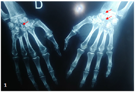
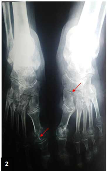
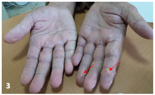
A 38-year-old man with a history of hyperuricemia for 1 year on Purinol300 1 tablet/day and hypothyroidism on replacement therapy was admitted for inflammatory polyarthralgia of the wrists and ankles. On investigation, the patient reported repeated acute gouty attacks in the right big toe. The clinical examination revealed a painful limitation of the wrists and ankles and a flessum of the left elbow. Biology showed a biological inflammatory syndrome, hyperuricemia at 664Umol/l and normal blood calcium/ phosphorus/creatinine. Rheumatoid factor and anti-CCP were negative. X-rays of the hands showed geodes of the carpus and resorption of the 3rd phalangeal hoop (Figure 1). X-rays of the feet revealed notches of the right 1st metatarsal with a “halberd” appearance, geodes of the right 5th metatarsal and tarsitis of the left foot (Figure 2). The diagnosis of polyarticular gouty arthropathy was accepted. The patient was put on colchicine and allopurinol in addition to the hygienic-dietary rules. The initial evolution was good with regression of the arthritis, but given the poor compliance with the treatment, the evolution was marked by iterative, more frequent and multi-articular gouty attacks affecting the metacarpophalangeal, proximal interphalangeal and metatarsophalangeal joints. He was operated on twice for suspected septic arthritis of the knee, for which bacteriological samples were negative. Hard, painless, whitish nodular lesions of varying size and irregular surface adherent to the deep plane (Figure 3) appeared in the palms and elbows 10 years after the first attack of gout. These lesions were suggestive of gouty tophi. They were ulcerated in places, leaving a chalky sludge.
Figure 1: Hand X-ray: Geodes of the carpal bones + Notch of the large carpal bone.

Figure 2:Notches in the head of the right 1st metatarsal, “halberd” appearance Tarsal aspect of the left foot

Figure 3:Gouty tophi of the palmar surface of the hands.

Discussion
Gout is the most common intermittent inflammatory rheumatic disease in adult men after the age of forty. It is the consequence of chronic hyperuricemia and initially monoarticular sodium urate crystal deposits manifesting as acute recurrent painful arthritic attacks in the first metatarsophalangeal joint (MTP1). At the stage of tophaceous gout, these urate crystal deposits become ubiquitous and lead to chronic arthropathy and gouty tophi [1,2]. Our patient was presented with joint destruction particularly in the carpals and feet.
Tophaceous gout usually occurs more than 10 years after the initial presentation of the disease. Tophus is the pathognomonic feature of tophaceous gout. It consists of subcutaneous deposits of urate in the form of yellowish, firm, bulky subcutaneous nodules raised from thinned epidermis, preferentially located in the small distal joints of the hands and feet, elbows, knees, Achilles tendon and auricular cartilages [3,4]. Histologically, it is a chronic granulomatous inflammatory response to monosodium urate crystals that infiltrate the joints and soft tissues causing structural joint damage and disabling pain [1].
Tophaceous gout is the cause of significant structural joint damage including cartilage, bone and tendon. Several studies have demonstrated the link between the presence of intraosseous tophi and the occurrence of bone erosions, osteophytosis, cartilage destruction closely related to the sites of tophus deposition [5,6].
Our case shares some similarities with those of Aradoini et al. [7] in which he reported a case of severe chronic tophaceous gout with destructive joint involvement and large diffuse skin tophi in a 67-year-old man with an 8-years history of untreated gout [7].
The diagnosis of chronic tophoidal gout was made in our patient after excluding the diagnosis of rheumatoid arthritis due to the history of acute mono-articular gouty attacks in the big toe, the progression to the polyarticular destructive form, the appearance of gouty tophi, hyperuricemia, and the good initial response to colchicine and well-conducted hypouricemic therapy. Hypothyroidism was described as a pathological condition favoring the occurrence of gouty disease [8].
The treatment of tophaceous gout is based on background hypouricemic treatment with a xanthine oxidase inhibitor in the first line by allopurinol in combination with a hypouricemic diet targeting a uricemia lower than 50mg/L (300Umol/L). Theoretically, treatment options for the crisis include colchicine, NSAIDs, corticosteroids and IL-1 inhibitors for refractory cases [9,10]. Our observation is remarkable for its early age of onset, the location of uratic deposits in the palms of the hands, a location rarely described in the literature [3] and the severity of the destructive joint involvement. Only a few cases of erosive polyarticular gout have been identified in the literature [11].
Conclusion
This observation illustrates a severe form of tophaceous gout with joint involvement and the presence of subcutaneous tophi. This form presents a significant functional impact and highlights the importance of early and rigorous management of gouty disease to avoid or slow its progression and improve the patient’s quality of life..
Authors Contribution
*I Chabchoub and R Ben Salah: Collected data and information and were the major writers of the present work.
*O Frikha: Contributed to manuscript writing.
*F Frikha, S Marzouk, Z Bahloul: Contributed to data analysis and manuscript writing.
References
- Dalbeth N, Merriman TR, Stamp LK (2016) Gout. Lancet 388(10055): 2039-2052.
- Carey Berner I, Dudler J (2006) Rhumatologie La goutte. Rev Med Suisse 2: 30888
- Benmously Mlika R, Fenniche S, Marrak H, Badri T, Ben Jennet S, et al. (2006) Tophus goutteux. Ann Dermatol Venereol 133: 831-832.
- Kawtar Inani, Fatimazahra Merniss (2014) Le tophus goutteux. Pan African Medical Journal 17: 250.
- Popovich I, Dalbeth N, Doyle A, Reeves Q, McQueen FM (2014) Exploring cartilage damage in gout using 3-T MRI: Distribution and associations with joint inflammation and tophus deposition. Skeletal Radiol 43(7): 917-924.
- Dalbeth N, Aati O, Kalluru R, Gamble GD, Horne A, et al. (2014) Relationship between structural joint damage and urate deposition in gout: A plain radiography and dual-energy CT study. Ann Rheum Dis 74(6): 1030-1036.
- Aradoini N, Talbi S, Berrada K, Abourazzak FZ, Harzy T (2015) Chronic tophaceous gout with unusual large tophi: Case report. Pan Afr Med J 22: 132.
- Jean-Michel Boileau (2021) The treatment of gout. Blue Pages.
- Augustin Latourtea, Tristan Pascartb, René-Marc Flipoc, Gérard Chalèsd, Laurence Coblentz-Baumanne, et al. (2020) Recommendations of the French Society of Rheumatology for the management of gout: Management of acute flares. Joint Bone Spine 87(5): 387-393.
- Richette P, Doherty M, Pascual E, Barskova V, Becce F, et al. (2017) 2016 updated EULAR evidence-based recommendations for the management of gout. Ann Rheum Dis 76(1): 29-42.
- Aurelian Anghelescu (2019) Multiarticular deforming and erosive tophaceous gout with severe comorbidities. Journal of Clinical Rheumatology 26(7): e269-e271.
© 2023 Imen Chabchoub. This is an open access article distributed under the terms of the Creative Commons Attribution License , which permits unrestricted use, distribution, and build upon your work non-commercially.
 a Creative Commons Attribution 4.0 International License. Based on a work at www.crimsonpublishers.com.
Best viewed in


.jpg)


 Editorial Board Registrations
 Submit your Article
 Refer a Friend
 Advertise With Us
.jpg)


.jpg)


.bmp)
.jpg)
.png)
.jpg)


.jpg)


.png)

.png)


.png)


