- Submissions

Full Text
Advancements in Case Studies
The Effects of Diamagnetic Therapy in a Peripheral Facial Paralysis: A Case Report
Estefania Torres Sanchez1, Felipe Torres Obando2, Federica di Pardo3 and Pietro Romeo3*
1Faculty of Medicine and Health Sciences, University del Rosario, Colombia
2Faculty of Medicine and Health Sciences, University del Rosario-Cell Regeneration Medical Organization, Colombia
3Periso Academy, Lugano, Switzerland
*Corresponding author: Pietro Romeo, Periso Academy, Lugano, Switzerland
Submission:December 28, 2022;Published: January 10, 2023

ISSN 2639-0531Volume3 Issue5
Abstract
Peripheral facial paresis, or paralysis, is the most common acute mononeuropathy that affects the face. Also known as Bell’s paralysis and it involves the VII cranial nerve on one side of the face. The clinical resolution is almost always spontaneous, in a few weeks, but the complete recovery usually occurs in about six months; rarely, the pathology continues with long symptoms for life or occurs more than once. Despite the cause is frequently unknown, the therapeutic choice includes corticosteroids or antiviral drugs in force of a possible inflammatory or viral origin. In addition to medical therapy, short electrical stimulation of the facial muscles, low-level facial lasers, facial exercises, and tape feedback have been proposed, but with conflicting results.
We are unaware of the use of Pulsed Electromagnetic Fields in the management of paralysis, although this type of biophysical stimulation has already proven to be effective in neurological diseases, both in the central and peripheral nervous contexts. Based on this rationale, an original technology that employs High-Intensity Magnetic Fields (Diamagnetic therapy) has been proposed as a therapeutic choice in facial palsy given the lengthy response after conventional rehabilitative treatments. The results of the treatment are very encouraging and give way to future applications, also extended to other diseases of the peripheral nervous system.
Keywords:Facial paralysis; Bell’s paralysis; Pulsed electromagnetic fields; Diamagnetic therapy
Introduction
The incidence of peripheral facial paralysis is around 20 to 25 cases per 100.000 population annually and the age range that predominates in this pathology is between 15 to 45 years old people [1]. The cause can be idiopathic, infectious, traumatic, iatrogenic, or resulting from tumors or developmental abnormalities in the nerve, as part of isolate syndromes or traumatic birth injuries and early infections [2]. This condition affects the lower motor neuron tract on the face, specifically the cranial nerve VII, which has both motor and sensory components. Additionally, the sensory root of the facial nerve carries the parasympathetic innervation of some salivary, lacrimal and mucosa glands and the motor fibers supply the muscles of facial ex-pression, auricle and scalp [3]. The course of the facial nerve is one of the most complex compared to the other cranial nerves and comprises intracranial, infratemporal and extratemporal parts whose knowledge is crucial to locate and treating possible lesions by using biophysical stimulation of the nerve [4].
Bell’s palsy is characterized by abrupt impaired facial expression due to unilateral facial weakness and involves all facial nerve branches, which directly implies a dry eye due to the impossibility of closing the eyelids, posterior auricular pain, numbness, altered taste and decrease tearing [5]. The onset generally occurs between 48 to 72 hours after the cause: infections, anatomic anomalies, inflammation, cold exposure, and ischemia [6]. The main current hypotheses on the pathophysiology of Bell’s palsy are the reactivation of Herpes Simplex virus infection or other types of cell-mediated autoimmune inflammatory response. These episodes can be triggered by stress or an infection of other illnesses capable of triggering a reactive response against the myelin antigens of the facial nerve. In the pathogenesis, of the disease, inflammation edema, and swelling lead to nerve compression at the origin of the starting symptoms [7].
To define the presence of facial palsy a detailed examination of the patient’s clinical history and a physical examination are recommended. The american academy of otolaryngology-head and neck surgery does not recommend laboratory, electrodiagnostic or radio-logic testing or diagnostic imaging for the new onset of Bell’s palsy. However, in presence of red flags or suspects of other pathology, is necessary to derogate from this rule and look for other causes of the disorder [8]. The common treatment includes the use of corticosteroids within 72h of facial paralysis onset, physiotherapy that includes beat therapy, electrostimulation, massage, mime therapy and biofeedback [5] also alternative therapies such as acupuncture, homeopathic or vitamins [9].
The prognosis of this condition is characterized by complete recovery in about 80% of the cases, 15% experience some permanent nerve damage, and 5% of them remain with sequelae [4]. Patients who have not gotten a recovery in the first 3 or 4 months after onset are more likely to have an incomplete facial function [10]. Despite the well-known impacts of electromagnetic fields in nervous tissues, few experiences report on the effects of the external stimulation of the facial nerve as a possible therapeutic choice. On these bases, we report the clinical case of a patient with facial palsy treat-ed using an original technology based on the low frequency- high-intensity pulsed electromagnetic fields (Diamagnetic Therapy), as an add-on treatment.
Case Presentation
A female patient, 49 years old, suffering from an episode of peripheral facial paralysis because of a strong moment of stress in her work life, went to our observation at the Cell Regeneration Medical Organization in Bogotá -Colombia- to undergo rehabilitative and medical treatments.
The woman, within her recovery process, had been treated by professionals in neurology and physiotherapy, covering for approximately 4 months the standard procedures related to peripheral facial palsy (corticoids, mime therapy, acupuncture, biofeedback). Due to the slow functional recovery, we decided to treat the patient with an innovative technology base on the repulsive effects of the low frequency -high-intensity magnetic field (Diamagnetic Therapy), already experienced to accelerate the biological regenerative processes in an experimental and clinical setting [11,12], besides the general well-known anti-inflammatory and modulatory effects of magnetic fields in nerves [13]. To evaluate the outcomes of the treatment, the Facial Nerve Grading System (FNGS) has been employed pre- and post-treatment [14] while the Visual Analogical Score was used for pain evaluation.
Methods
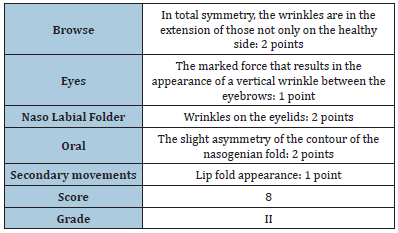
The preliminary clinical evaluation addressed the motor aspect of the disease. So, the ability to activate and move the facial muscles has been assessed and carried out before the first session of Diamagnetic treatment as follows (Table 1).
Table 1: Pain: According to VAS (Visual Analogic Score) 4/10.

Method of treatment
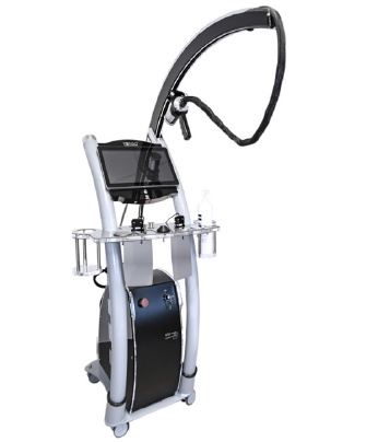
The Patient took 4 sessions, 1 per week, of diamagnetic therapy that includes high-intensity pulsed electromagnetic fields (2.2 Tesla), pulsed at low frequencies (7hz), named CTU Mega 20, provided by PERISO SA – Pazzallo (Switzerland) (Figure 1).
Figure 1: Diamagnetic Pump–CTU Mega 20 Device.

Protocol used for facial palsy
.The treatment was divided into 3 phases:
A. Phase Pain Control: 5 minutes neuropathic pain setting,
us-ing a mid-energy value due to the pain referred to in the
frontal muscle (frontalis muscle occipitofrontalis): VAS 4/10 on
a visual analog scale.
B. Second phase Endogenous Bio-Stimulation: 5 minutes
“nerve fast” over the facial nerve of the right hemiface.
C. 5 minutes “nerve slow” over the facial nerve of the right
hemi-face.
D. 5 minutes “skeletal muscle” over all the right hemiface
mus-cles.
E. Third phase movements of liquids: 5 minutes intracellular
and 5 minutes extracellular.
For a global time of 25 minutes treatment. The Diamagnetic treatment has been applied in the extracranial area at the emergence of the facial nerve. 50% of the treatment time was addressed to the correspondence of the geniculate nerve ganglion and the remaining part was to treat the ophthalmic, maxillary, and mandibular areas working in static and dynamic modes, with different times.
Results
Muscle movement evaluation: fourth session (FNGS score) (Table 2).
Table 2: Pain, after the fourth session 1/10 according to the VAS score.

Discussion
Facial nerve palsy causes a lack of facial expression and loss of functional capacities, resulting in movement difficulties, visual impairments and oral incompetence. These limitations have a big impact on social life and the patients affected may experience anxiety and depression, also as a consequence of a decreased quality of life [15]. Early diagnosis and treatment are mandatory, also considering that a delay can affect the long-term results. Both surgical and nonsurgical treatments are commonly performed. However, in the case of Bell’s palsy, the nerve is anatomically intact and before surgery, a period of observation is recommended. It is not clear how long this period should be, but generally, a lack of improvements after 6 months of observation predicts no satisfactory long-term recovery [16]. A recent Cochrane systematic review confirms that there is no certainty on surgical intervention within three months [17] and up to 85% of patients with Bell’s palsy treated with placebo report a complete recovery within 1 year. Those with remaining facial weakness, especially of eyelid muscles, could be candidates for surgery.
According to the American Academy of Neurology (AAN), in the case of Bell’s palsy, it is recommended with a level A the use of cortico-steroids within 72 hours of onset. The addition of antiviral drugs does not offer significant benefits, but some studies could not rule out a possible effect and, for this reason, in general practice antivirals are often used [18]. Non-surgical treatments of facial palsy also include specific rehabilitative therapy. It consists of 5 main components: patient education, soft tissue mobilization, functional retraining concerning oral abilities, facial expression and synkinesis management [19]. The role of physical therapy like brief electrical stimulation of facial muscles, low-level facial laser, gross facial exercises and tape feedback are still controversial [20]. To our knowledge, no author has ever investigated the efficacy of electromagnetic field stimulation for facial palsy recovery. However, there is proof of evidence on the effects of magnetic fields once applied for the treatment of peripheral neurological pathologies in force of re-generative and ant-inflammatory effects [21,22]. Moreover, in the past transcranial magnetic stimulation (TMS) and peripheral stimulation of cranial nerves were investigated as methods of functional assessment of the rapid conducting corticobulbar and corticospinal projections in brainstem lesions [23] as well as the excitation of cortico-facial fibers at the brainstem level and the TMS at the posterolateral scalp to stimulate the nerve at the facial canal region or the labyrinthine segment of the nerve [24]. This approach derives from positive results by stimulating cranial nerves in the course of neuropsychiatric disorders, including the triggering of neuronal plasticity and potentiating synaptic transmission [25].
The technology we employed in our experience is quite different from other types of nervous stimulation. Specifically, we used a pulsed high-intensity low-frequency electromagnetic field (PEMF) delivered by the Diamagnetic Pump device (CTU Mega 20). This technology allows a treatment called diamagnetic therapy, which is safe for the patient thanks to the low frequency of the magnetic field (<50 Hz) and belongs to the non–ionizing radiation class. On the other hand, the high intensity (up to 2 Tesla) guarantees a diamagnetic effect on the treated tissues, consisting of a repulsive action (movement) of the water, solutes, ions and molecules, able to improve the electrochemical changes at the cell membrane level that is at the origin of the biological response in the treated tissues [11]. The intrinsic frequency of the magnetic field is 7500Hz, and the duration of the pulses is 5ms with a period of 1000ms, a wide bandwidth of the electromagnetic frequencies guarantees the possibility to treat selectively in the same session nerves, muscle and bone [26]. In the neurological field, the technology has been analyzed in a randomized double-blind sham-controlled trial which has shown that the non-ionizing LF-PEMFs induced by the CTU Mega 20 cause an increase of more than 60% in corticospinal excitability in healthy subjects [27], also showing behavioral changes and qualitative improvements in a case series of rare-orphan diseases [28].
Given the above-mentioned prerogatives of Diamagnetic therapy, characterized also by the possibility to modulate the rise time of the Magnetic Pulse, the amplitude and the bandwidth of the electro-magnetic frequencies, it was possible to treat selectively, the ganglion, the muscles and nerve fibers both in static and dynamic mode. In this way, the symptoms, the clinical evidence and the changes reported during the treatment addressed the machine’s setting. We started to modulate in order: the referred pain in the frontal region (VAS 4/10 at the beginning), the course of the facial nerve using selected frequencies for fast and slow nerve fibers, for muscles and to promote the extra and intracellular movement of liquids, ions and molecules. 50% of the treatment time was addressed to the correspondence of the geniculate nerve ganglion and the remaining part was to treat the ophthalmic, maxillary and mandibular areas working in static and dynamic mode, but not in the equivalent distribution of the time between the three branches of the facial nerve. This is due to the greater suffering of the mandibular branch. These positive effects would be related to the modulation of the inflammatory edema and the swelling that normally lead to nerve compression, given the repulsive effect on liquids induced by the high-intensity magnetic field [27], together with the selected stimulation of the nerve fibers.
We are aware of the weakness of a single case report without an equivalent case control but, as often happens, a single clinical report acts as a driving force for further studies with a suitable statistical sample and in a randomized and controlled form. Anyway, the results underline the fast efficacy of the treatment after four weekly sessions, as shown by the changes in the functional Facial Nerve Grading System (from Grade V to Grade II) and the fast recovery in pain (VAS 4/10 to 1/10). These last states a difference > of 2 points and, consequently, higher respect to the MCID (Minimally Clinically Important Difference).
Conclusion
Diamagnetic Therapy is a versatile ready-to-use technology that, according to the PEMF prerogatives allows, in an original way, effective and selected treatments in the different tissues including the nervous system. Further studies are necessary to valorize the results of our experience, and to exploit the therapy in other pathological conditions of the nerves. Moreover, this innovative technology may be tested for the release of heat from the low frequency high-intensity magnetic field, since heat may inactivate the heat shock gene Sirtuin 1 which is critical for neuroplasticity and neural stem cell regeneration [29,30], also considering that this is controversial with the absence of thermal effects observed, in terms of HSP90 expression, in different cell experimental settings employing Low-Frequency Magnetic Field ( < 50Hz) [31].
References
- Finsterer Jose (2008) Management of peripheral facial nerve palsy. Eur Arch Otorhinolaryngology 265(7): 743-752.
- Owusu JA, Stewart CM, Boahene K (2018) Facial Nerve Paralysis. Medical Clinics of North America 102(6): 1135-1143.
- Lorch M, Teach SJ (2010) Facial nerve palsy: Etiology and approach to diagnosis and treatment. Pediatric Emergency Care 26(10): 763-769.
- Yang SH, Park H, Yoo DS, Joo W, Rhoton A (2021) Microsurgical anatomy of the facial nerve. Clinical Anatomy 34(1): 90-102.
- Jimmy HO (2022) Diagnosis and management of bell’s palsy in primary care. The Journal for nurse practitioners 18(2): 159-163.
- Zhang W, Xu L, Luo T, Wu F, Zhao B, et al. (2020) The etiology of bell´s palsy: A review. Journal of Neurology 267(7): 1896-1905.
- Heckmann JG, Urban PP, Pitz S, Guntinas-Lichius O, Gagyor I (2019) The diagnosis and treatment of idiopathic facial paresis (Bell’s Palsy). Dtsch Arztebl Int 116(41): 692-702.
- Kim SJ, Lee HY (2020) Acute peripheral facial palsy: Recent guidelines and a systematic review of the literature. J Korean Med Sci 35(30): e245.
- Lassaletta L, Morales-Puebla JM, Altuna X, Arbizu A, Aristegui M, et al. (2019) Facial paralysis: a clinical practice guideline of the Spanish society of otolaryngology. Acta Otorrinolaringol Esp 71(2): 99-118.
- Timothy J, Croxson GR, Kennedy PGE, Hadlock T, Krishnan AV (2015) Bell’s palsy: aetiology, clinical features and multidisciplinary care. J Neurol Neurosurg Psychiatry 86(12): 1356-1361.
- Carnovali M, Stefanetti N, Galluzzo A, Romeo P, Mariotti M, et al. (2022) High-intensity low-frequency pulsed electromagnetic fields treatment stimulates fin regeneration in adult zebrafish-a preliminary report. Appl Sci 12(15): 7768.
- Roberti R, Marcianò G, Casarella A, Rania V, Palleria C, et al. (2022) High-intensity, low-frequency pulsed electromagnetic field as an odd treatment in a patient with mixed foot ulcer: A case report. Reports 5(1): 3.
- Kanjanapanang N, Chang KV (2022) Peripheral magnetic stimulation. In: Stat Pearls [Internet]. Treasure Island (FL): Stat Pearls Publishing.
- Mengi E, Kara CO, Ardıç FN, Barlay F, Çil T, et al. (2020) Validation of the Turkish version of the facial nerve grading system 2.0. Turk Arch Otorhinolaryngology 58(2): 106-111.
- Robinson MW, Baiungo J (2018) Facial rehabilitation. Otolaryngologic Clinics of North America 51(6): 1151-1167.
- Owusu JA, Stewart CM, Boahene K (2018) Facial nerve paralysis. Medical Clinics of North America 102(6): 1135-1143.
- Menchetti I, McAllister K, Walker D, Donnan PT (2021) Surgical interventions for the early management of Bell’s palsy. Cochrane Database of Systematic Reviews. Edited by Cochrane Neuromuscular Group.
- Reich SG (2017) Bell’s palsy. Continuum: Lifelong Learning in Neurology 23(2): 447-466.
- Robinson MW, Baiungo J (2018) Facial rehabilitation: Evaluation and treatment strategies for the patient with facial palsy. Otolaryngologic Clinics of North America 51(6): 1151-1167.
- Van Landingham SW, Diels J, Lucarelli MJ (2018) Physical therapy for facial nerve palsy: Applications for the physician. Current Opinion in Ophthalmology 29(5): 469-475.
- Abdi S (2018) Treatment of chemotherapy-induced peripheral neuropathy: Systematic review and recommendations. Pain Physician 21(6): 571-592.
- Liampas A, Rekatsina M, Vadalouca A, Paladini A, Varrassi G, et al. (2020) Non-Pharmacological management of painful peripheral neuropathies: A systematic review. Advances in Therapy 37(10): 4096–4106.
- Urban PP (2003) Chapter 35 Transcranial magnetic stimulation in brainstem lesions and lesions of the cranial nerves. Supplements to Clinical Neurophysiology 56: 341-357.
- Lo YL, Fook-Chong S (2007) Magnetic brainstem stimulation of the facial nerve. Journal of Clinical Neurophysiology 24(1): 44-47.
- Iglesias AH (2020) Transcranial magnetic stimulation as treatment in multiple neurologic conditions. Current Neurology and Neuroscience Reports 20(1): 1.
- Obando AFT, Velasco JM, Romeo P (2020) Variable low frequency-high intensity-pulsed electromagnetic fields in the treatment of low back pain: A case series report and a review of the literature. J Orthop Res Ther 5(4): 5.
- Premi, E, Benussi A, La Gatta A, Visconti S, Costa A, et al. (2018) Modulation of long-term potentiation-like cortical plasticity in the healthy brain with low frequency-pulsed electromagnetic fields. BMC Neuroscience 19(1): 34.
- Obando FT, Romeo P, Vergara D, Di Pardo F, Soto A (2022) The effects of low-frequency high-intensity pulsed electromagnetic fields (diamagnetic therapy) in the treatment of rare diseases: A case series preliminary study. J Neurol Exp Neural Sci 4: 145.
- Ian James Martin (2019) Human survival and immune mediated mitophagy in neuroplasticity disorders. Neural Regeneration Research 14(4): 735.
- Alexzander A, Asea A, Kaur P (2019) Heat shock proteins in neuro-science. Immune Reactions and Species Longevity, Volume 20.
- García-Minguillán O, Prous R, Del Carmen Ramirez-Castillejo M, Maestú C (2020) CT2A cell viability modulated by electromagnetic fields at extremely low frequency under no thermal effects. Int J Mol Sci 21(1): 152.
© 2023 Pietro Romeo. This is an open access article distributed under the terms of the Creative Commons Attribution License , which permits unrestricted use, distribution, and build upon your work non-commercially.
 a Creative Commons Attribution 4.0 International License. Based on a work at www.crimsonpublishers.com.
Best viewed in


.jpg)


 Editorial Board Registrations
 Submit your Article
 Refer a Friend
 Advertise With Us
.jpg)


.jpg)


.bmp)
.jpg)
.png)
.jpg)


.jpg)


.png)

.png)


.png)


