- Submissions

Full Text
Forensic Science & Addiction Research
Principles of Polarization Tomography Multifunctional Digital Histology in the Diagnosis of Prescription Damage to Internal Organs
Alexandra Litvinenko1 and Oleg Vanchulyak2*
1Postgraduate student of the Department of Forensic Medicine and Medical Law, Bukovinian State Medical University, Ukraine
2Professor of the Department of Forensic Medicine and Medical Law, Bukovinian State Medical University, Ukraine
*Corresponding author: Oleg Vanchulyak, Professor of the Department of Forensic Medicine and Medical Law, Bukovinian State Medical University, Chernivtsi, Ukraine
Submission: June 09, 2022;Published: July 07, 2022

ISSN 2578-0042 Volume5 Issue5
Abstract
For high-precision objective histological determination of the prescription of damage to internal organs over a long period of time, a systematic approach was used based on digital azimuthally invariant polarization, Mueller-matrix and tomographic methods for studying temporal changes in the molecular and polycrystalline structure of brain, liver and kidney samples in the postmortem period. It was revealed that the linear change in the magnitude of statistical moments of the 1st - 4th orders, characterizing the distribution of data from digital azimuthally invariant polarization, Muller-matrix and tomographic methods, is interconnected with the duration of damage to internal organs in the time interval from 1 hour up to 120 hours. On this basis, a new algorithm for digital histological determination of the age of damage is proposed.
Keywords: Histology; Polarization; Mueller matrix; Tomography; Statistical moments; Determining the age of damage
Highlights
The main idea of our study is based on the existence of diagnostically important relationships between traditional microscopic images, as well as computer polarized and tomographic images new to histological practice, and quantitative (digital) characteristics of their topographic maps. The study of the diagnostic capabilities of multiparametric digital histology is based on the complex application of polarization [1], Mueller-matrix [2- 4], and tomographic [5-7] mapping techniques and the use of objective statistical analysis of the obtained microscopic and computer images. Certain parameters of such an analysis (statistical moments of the 1st - 4th orders [8]) are the basis for establishing objective forensic criteria for determining the prescription of damage to internal organs. The design of our study is illustrated by the following structural and logical diagram, - Table 1.
Table 1: The design of our study.

Materials and Methods
Polarization azimuth maps of microscopic images of histological sections of biological tissues of human internal organs
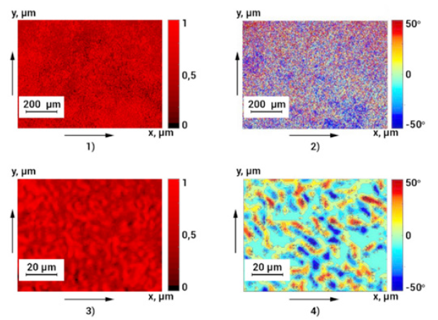
This part presents a comparative presentation of the topographic structure of traditional microscopic images (4x and 40x magnification) of a histological section of the brain that died due to Coronary Heart Disease (CHD) (Figure 1, fragments ((1),(3))) and the corresponding maps of polarization azimuth such images (Figure 1, fragments ((2),(4)). It can be seen from the data obtained (Figure 1) that, along with the topographic structure of the intensity values of traditional microscopic images ((1), (3)) their polarization maps are also more clearly structural ((2), (4)) as the spread of the azimuth values polarization, and by its average level. This result indicates additional information reserves for studying computer polarization maps of such images in order to obtain new information about changes in the biochemical and molecular structure of samples of biological tissues of human internal organs with different damage duration. Note that the above example of polarization mapping of azimuth and polarization is related to the microscopic image of a biological preparation itself and does not carry information about its direct morphological, biochemical and molecular structure. Such data can be obtained using other polarization techniques - Mueller-matrix mapping [6].
Figure 1: Microscopic images ((1), (3)) and polarization azimuth maps ((2), (4)) of a histological section of the brain of a person who died from CHD.

Method of Mueller-matrix mapping of histological sections of biological tissues of human internal organs
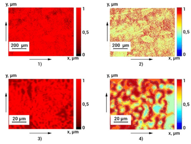
In a series of fragments in Figure 2 shows microscopic images ((1),(3)) and maps of Mueller-matrix images ((2),(4)) of linear birefringence (degree of crystallization) of the liver of a person who died of CHD. Analysis of certain (Figure 2) maps of Muller-matrix images of linear birefringence ((2),(4)) revealed a significant structural heterogeneity in the degree of crystallization of the histological section of the liver compared to the topographic structure of the magnitude of the intensity of traditional microscopic images ((1),(3)). Azimuthally invariant Muller-matrix mapping of the crystal and molecular structure of histological sections of biological tissues of internal organs provides the possibility of a comprehensive analysis of changes in the structure of their substance as a result of traumatic injuries. Note that the above examples of polarization and Muller-matrix mapping are related to indirect information about the structure of biological objects and their microscopic images. Therefore, it is important from an applied (diagnostic) point of view to supplement the considered methods with the latest approaches of polarization tomography, which allow reconstructing maps of optical anisotropy (birefringence and optical activity) of histological sections of biological tissues and films of biological fluids.
Figure 2: Microscopic images ((1), (3)) and Mueller-matrix images of linear birefringence ((2), (4)) of a histological section of the liver of a deceased from CHD.

The technique of polarization reconstruction (tomography) of the polycrystalline structure of histological sections of biological tissues of human internal organs
The technique for reconstructing the parameters of the polycrystalline component (linear and circular birefringence) of the substance of tissues of human internal organs is presented in detail in scientific papers [5-7]. It is based on successive probing of a biological preparation with differently polarized light beams, polarization filtering of a series of microscopic images, and algorithmic reconstruction of the coordinate distributions of the magnitude of linear and circular birefringence. On the fragments of Figure 3 shows examples of polarization reconstruction of the degree of crystallization of the substance of a histological section of a kidney of a person who died from CHD. Comparison (Figure 3) of traditional microscopic images ((1),(3)) and tomographically reproduced maps of linear birefringence of fibrillar structures ((2),(4)) of a histological section sample revealed a significant range of change and coordinate heterogeneity in the degree of tissue crystallization. The information obtained in this way allows direct detection (inaccessible to traditional light microscopy methods) and quantitative assessment of changes in the spatial structure of fibrillar networks of the morphological structure of tissues of human internal organs due to traumatic injuries of various prescriptions..
Figure 3:Microscopic images ((1), (3)) and maps of linear birefringence ((2), (4)) of a histological section of a kidney of a person who died of CHD.

Conclusion
1. The most sensitive parameters and the following ranges
of linear change in the magnitude of statistical indicators of
polarization digital histology and the accuracy of determining the
age of damage were identified:
A. Polarization azimuth maps of microscopic images
(asymmetry, kurtosis – 12 hours, accuracy – 55 min. - 60 min.);
B. Polarization ellipticity maps of microscopic images
(asymmetry, kurtosis – 12 hours, accuracy – 65 60 min. – 75 60
min.).
2. Diagnostically sensitive parameters have been established
- time ranges of linear change in the magnitude of statistical
indicators of Muller-matrix digital histology methods and the
accuracy of determining the age of damage:
A. MMI maps of the degree of crystallization (asymmetry,
kurtosis – 24 hours, accuracy – 55 min. – 60 min.);
B. MMI maps of optical activity (asymmetry, kurtosis – 24
hours, accuracy – 65 min. – 75 min.).
3. A set of time ranges for linear changes in the magnitude of
statistical moments of 1-4 orders of magnitude characterizing the
distribution of data of the tomographic technique of optical activity
of molecular complexes of digital histology and the accuracy of
determining the duration of damage to internal organs of a person
has been established:
A. Average – 72 hours, accuracy 30 min. – 35 min.
B. Dispersion – 72 hours, accuracy 30 min. – 35 min.
C. Asymmetry – 120 hours, accuracy 15 min. – 25 min.
D. Kurtosis – 120 hours, accuracy 15 min. – 25 min.
References
- Ushenko VA, Hogan BT, Dubolazov A, Grechina AV, Boronikhina TV, et al. (2021) Embossed topographic depolarisation maps of biological tissues with different morphological structures. Sci Rep 11(1): 3871.
- Trifonyuk L, Sdobnov A, Baranowski W, Ushenko V, Olar O, et al. (2020) Differential Mueller matrix imaging of partially depolarizing optically anisotropic biological tissues. Lasers in Medical Science 35(4): 877-891.
- Ushenko VA, Hogan BT, Dubolazov A, Piavchenko G, Kuznetsov SL, et al. (2021) 3D Mueller matrix mapping of layered distributions of depolarisation degree for analysis of prostate adenoma and carcinoma diffuse tissues. Scientific Reports 11(1): 5162.
- Borovkova M, Trifonyuk L, Ushenko V, Dubolazov O, Vanchulyak O, et al. (2019) Mueller-matrix-based polarization imaging and quantitative assessment of optically anisotropic polycrystalline networks. PLoS ONE 14(5): e0214494.
- Ushenko V, Sdobnov A, Syvokorovskaya A, Dubolazov A, Vanchulyak O, et al. (2018) 3D Mueller-matrix diffusive tomography of polycrystalline blood films for cancer diagnosis. Photonics 5(4): 54.
- Peyvasteh M, Tryfonyuk L, Ushenko V, Syvokorovskaya AV, Dubolazov A, et al. (2020) 3D Mueller-matrix-based azimuthal invariant tomography of polycrystalline structure within benign and malignant soft-tissue tumours. Laser Physics Letters 17(11): 115606.
- Borovkova M, Peyvasteh M, Dubolazov O, Ushenko Y, Ushenko V, et al. (2018) Complementary analysis of Mueller-matrix images of optically anisotropic highly scattering biological tissues. Journal of the European Optical Society 14(1): 20.
- Sarkisova Y, Bachinskyi VT, Garazdyuk M, Vanchulyak OY, Litvinenko OY, et al. (2020) Differential muller-matrix microscopy of protein fractions of vitreous preparations in diagnostics of the pressure of death. IFMBE Proceedings 77: 503-506.
© 2022 Oleg Vanchulyak. This is an open access article distributed under the terms of the Creative Commons Attribution License , which permits unrestricted use, distribution, and build upon your work non-commercially.
 a Creative Commons Attribution 4.0 International License. Based on a work at www.crimsonpublishers.com.
Best viewed in


.jpg)


 Editorial Board Registrations
 Submit your Article
 Refer a Friend
 Advertise With Us
.jpg)


.jpg)


.bmp)
.jpg)
.png)
.jpg)


.png)

.png)


.png)


