- Submissions

Full Text
Surgical Medicine Open Access Journal
Non Tuberculous Mycobacterium as a Causative Factor in Port Site Wound Infection - A Case Report
JS Rajkumar*, Aishwarya Vinoth, S Akbar, Anirudh Rajkumar, Hema Tadimari, Aluru Jayakrishna Reddy and Shreya Rajkumar
Department of Surgery, SRM University, India
*Corresponding author: JS Rajkumar, Department of Surgery, SRM University, India
Submission: December 11, 2017; Published: January 19, 2018

ISSN: 2578-0379Volume1 Issue3
Abstract
Over the two decades there has been an increase in Non Tuberculous Mycobacterium (NTM) organisms in post laparoscopic port site skin and soft tissue infections. Presenting a patient who underwent eight successive surgical explorations of multiple skin and soft tissue sinuses and for aggressive necrosectomy and debridement of multiple sinuses caused by NTM organisms which ended up in abdominoplasty and meshplasty 2 months after the infection had been completely controlled. She finally had complete relief from the infection and had 6 months follow up from the last meshplasty surgery. The presentation and treatment of NTM skin and soft tissue infection are briefly outlined.
Keywords: Tuberculous; Mycobacterium; Infection; Meshplasty
Abbreviations: NTM: Non Tuberculous Mycobacterium; AFB: Acid Fast Bacilli; GTA: Gluteraldehyde
Introduction
Over the two decades there has been an increase in Non Tuberculous Mycobacterium (NTM) organisms in post laparoscopic port site skin and soft tissue infections. Presenting a patient who underwent eight successive surgical explorations of multiple skin and soft tissue sinuses and for aggressive necrosectomy and debridement of multiple sinuses caused by NTM organisms which ended up in abdominoplasty and meshplasty 2 months after the infection had been completely controlled. She finally had complete relief from the infection and had 6 months follow up from the last meshplasty surgery. The presentation and treatment of NTM skin and soft tissue infection are briefly outlined
Case Report
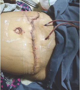
A 38 yr old lady presented to our institution with recurrent discharging sinuses over the anterior abdominal wall in relation to the ports of the laparoscopic ovarian cystectomy that she had undergone in 2013. Two wound excisions had been done by the primary surgeon and the sinus still recurred. Few weeks after the surgery patient developed abdominal pain and pus draining from the port site. In view of the pain and discharge, laparotomy was done and pus was drained out. Patient was started on antibiotics and in 2 weeks patient developed pustules and started draining pus. In another week the same pattern of redness, pustule and discharge started in 2 other areas. When patient came to us she had multiple pus discharging sinuses from the anterior abdominal wall which were closely related to the port site. Patient was otherwise stable and examination revealed severe abdominal wall tenderness with multiple discharging sinuses and regional lymphadenopathy in both groins. Patient was operated, fisulectomy was done and specimen sent for histopathology, pus sent for mycobacterial culture. Histopathology showed acute on chronic granulomatous inflammation. Acid Fast Bacilli (AFB) smear was negative for AFB bacilli and culture was positive for non mycobacterium tuberculosis rapid growers. Patient was started on inj. amikacin, clarithromycin, ofloxacin. During the course of the treatment, patient developed recurrent fistula and cultures were positive for MTB, for which patient was started on rifampicin, ethambutol, isoniazid, clarithromycin, pyrazinamide, and doxycycline. She was operated twice and every time the culture was sent and was positive for NTM. After 6 months of treatment the cultures were negative for NTM and MTB. Patient finally had abdominoplasty and mesh repair (Figure 1). Patient was continued on treatment for another 3 months. Now patient is not on treatment for 6 months, patient is stable, has no abdominal pain, fistula formation or wound site infection. Operative site is healthy and healed.
Discussion
Post laparoscopic surgery one can expect 2 types of infection; one within a week of surgery and another 3-4 weeks later which is mostly caused by NTM organisms [1]. In laparoscopic procedures the commonest site for infection is the umbilical port site and the second most commonly affected site is the epigastric port, from there it spreads to other port sites. The diagnosis is usually made from the tissue culture which takes about 3-4 weeks. So these days the early and newer diagnostic methods a such as BACTEC and RT- PCR is considered [1,2]. Clinical diagnosis is the best considered option. Port site infection is presented in 5 stages, 1st stage the patient develops a tender nodule on the port site. The 2nd stage the nodule becomes bigger and tender and starts discharging pus. The 3rd stage, it turns to be a sinus discharging pus. Stage 4, these develop into chronic pus discharging sinuses. 5th stage is the darkening of the necrotic skin [2]. If this infection is not treated with appropriate antibiotics it turns out to be a chronic pus discharging sinus with spread to adjacent areas and forms chains of sinuses.
Figure 1: Post abdominoplasty.

Laparoscopic instruments are also a source of infection. These instruments have a layer of insulation that restricts autoclaving [2]. So the infection spreads from the instruments and from the water used to wash laparoscopic instruments which is supplied from the over head tanks which are not cleaned properly. These are the source of NTM organisms like M.Marinum, M.Kansasii, M.Avium [3]. Laparoscopic instruments are usually cleaned with Gluteraldehyde (GTA), but atypical mycobacterium like M.Fortutium, Chelonae has showed resistance to these chemicals in humans. The endospores of this NTM complex are usually considered saprophytes which colonize in the soil and tap water. For the treatment of NTM, combination of first line ATT, Macrolides, Quinolones, Aminoglycolides, and Tetracyclines are used [4,5] .
To prevent post surgical infections, frequent cleaning of the instruments to be done. Endospores from contaminated instruments gets deposited in the subcutaneous tissue, which germinate in the tissue in about three to four weeks to produce clinical symptoms. Other liquid agents like orthophthaldehyde and per acetic acid can be replaced for GTA for higher levels of disinfection [5,6].
Recent studies have reported a significant increase in the port site infection with increase in preoperative stay of more than 2 days for open surgical procedures. They have also reported that there is no infection rate in surgeries less than 30min of duration. Port site infection are more common in the umbilical port [7,8]; the site through which the specimen is extracted is mostly commonly infected, so the infected specimen should be removed in an endobag in order to prevent wound infection and accidental spillage of contents or its contact over the skin and subcutaneous tissue [9].
Conclusion
In this case report, we have highlighted the importance of proper sterilization of laparoscopic instruments and cleaning of the over head tanks. This paper also highlights the importance of early diagnosis of NTM infections from its very specific characteristic clinical presentation in various stages since the cultures take a longer time and also the treatment regimen .So it is worth remembering that NTM organisms can be a major cause of port site wound infection and its complexity of treatment which can be avoided by maintaining good hospital hygiene.
References
- Vijayaraghavan R, Chandrasekar R, Sujatha Y, Belagavi CS (2006) Hospital outbreak of atypical mycobacterial infection of port sites after laparoscopic surgery: Journal of hospital infection. J Hosp Infect 64(4): 344-347.
- Katoch VM (2004) Infections due to non-tuberculous mycobacteria (NTM). Indian J Med Res 120: 290-304.
- Wallace RJ, O Brien R, Glassroth J, Raleigh J, Dutta A (1990) Diagnosis and treatment of disease caused by non-tuberculous mycobacteria. Am Rev Respir Dis 142: 940.
- Kalitha JB, Rahman H, et al. () Delayed post operative wound infections due to tuberculous mycobacterium.
- Min KW, Ko JY, Park CK, et al. (2012) Histopathological spectrum of cutaneous tuberculosis and non-tuberculous mycobacterial infections. J Cutan Pathol 39: 582-595.
- Dodiuk-Gad R, Dyachenko P, Ziv M, Shani-Adir A, Oren Y, et al. (2007) Nontuberculous mycobacterial infections of the skin: a retrospective study of 25 cases. Am Acad Dermatol 57(3): 413-420.
- Lilani SP, Jangale N, Chowdhary A, Daver GB (2005) Surgical site infection in clean and clean-contaminated cases. Indian J Med Microbiol 23(4): 249-252.
- Richards C, Edwards J, Culver D, Emori TG, Tolson J, et al. (2003) Does using a laparoscopic approach to cholecystectomy decrease the risk of surgical site infection? Ann Surg 237(3): 358-362.
- Taj MN, Iqbal Y, Akbar Z (2012) Frequency and prevention of laparoscopic port site infection. J Ayub Med Coll Abbottabad 24: 197-199.
© 2018 JS Rajkumar, et al. This is an open access article distributed under the terms of the Creative Commons Attribution License , which permits unrestricted use, distribution, and build upon your work non-commercially.
 a Creative Commons Attribution 4.0 International License. Based on a work at www.crimsonpublishers.com.
Best viewed in


.jpg)


 Editorial Board Registrations
 Submit your Article
 Refer a Friend
 Advertise With Us
.jpg)


.jpg)


.bmp)
.jpg)
.png)
.jpg)


.png)

.png)


.png)


