- Submissions
Full Text
Research in Pediatrics & Neonatology
Agenesis of the Corpus Callosum: Magnetic Resonance Imaging in Antenatal and Postnatal Cases
Markovic I1 and Milenkovic Z2*
1Institute of Radiology, Clinical Center Nis, Serbia
2General Hospital Sava Surgery, Serbia
*Corresponding author: Zoran Milenkovic, General Hospital “Sava Surgery”, Bvl. dr. Zoran Djindjic 91, Nis, Serbia, Email: zoran@ni.ac.rs
Submission: September 26, 2017; Published: November 13, 2017

ISSN : 2576-9200Volume1 Issue2
Abstract
Agenesis of the corpus callosum (ACC) is a birth defect manifested by the complete absence of the corpus callosum. The true incidence is dependent on chance because many isolated cases are asymptomatic. It is estimated that the frequency ranges between 0.5 and 70 in 10,000 birth or adult population and is evidently higher in pediatric cases with development anomalies (230 in 10,000 ) or it can reach 2-3 per 100 birth . Genetic factors are considered to be the main cause of ACC. There are no specific signs and symptoms of the malformation. Some of them maybe the result of other associated developmental abnormalities. In cases with isolated ACC, clinical syndromes are divers and vary in the severity of the clinical picture. Magnetic resonance imaging (MRI) is the procedure of choice and is a confirmative method in infants and children as well as in fetuses from about 20 weeks’ gestation. In our series of 4000 MRI explorations in children of different ages, 10 cases harboring ACC were diagnosed, accounting for 0.25%.On the other hand, in 48 pregnant women who were underwent MR investigation between 20 to 36 gestational weeks, another six cases with absence of the corpus callosum were diagnosed.
Keywords: Agenesis of corpus callosum; Epidemiology; Symptomatology; Magnetic resonance imaging
Abbrevations: ACC: Agenesis of the Corpus Callosum; MRI: Magnetic Resonance Imaging
Introduction
Agenesis of the corpus callosum (ACC) is a birth defect manifested by the complete absence of the corpus callosum, due to a disruption of the brain cell migration during the fetal development of a child. Although ACC can be an isolated disorder, it is frequently associated with other anomalies and neuro anatomical lesions such as the aneuploidy and non-aneuploidy syndromes, other CNS associations, as well as inborn errors of the metabolism [1]. Complete corpus callosum agenesis is a frequent anomaly. The true incidence is dependent on chance because many isolated cases are asymptomatic. It is estimated that the frequency ranges between 0.5 and 70 in 10,000 birth or adult population [2] and is evidently higher in pediatric cases with development anomalies (230 in 10,000 ) or it can reach 2-3 per 100 birth [3].
Genetic factors are considered to be the main cause of ACC. Several syndromes and several causative genes have been recognized [2]. Exogenic factors, such as atenatal infections, vascular or toxic insults, are involved less frequently in the development of the disorder. For isolated ACC an “interaction of a number of ‘modifier’ genetic and environmental factors” has been reported to be responsible for the occurrence of ACC [4]. There are no specific signs and symptoms of ACC. Some of them maybe the result of other associated developmental abnormalities. In cases with isolated ACC, clinical syndromes are divers and vary in the severity of the clinical picture. Clinical presentations are in this order of frequency: mental retardation, visual problems, speech delay, seizures, and feeding problems [5]. Other signs and symptoms have also been identified which include developmental delays in motor and language skills, vision and hearing impairment, poor muscle tone and coordination, insomnia or other sleep problems, psychosocial and cognitive difficulties. Individuals with mild symptomatology may have normal intelligence. Matteo Chiappedi et al. [6] described a case harboring the isolated ACC who has had mild learning difficulties with preserved intelligence.
MRI appearance of ACC
Magnetic resonance imaging (MRI) is the procedure of choice in the diagnosis of ACC. It is a confirmative method in infants and children as well as in fetuses from about 20 weeks’ gestation, possessing the significant sensitivity for illustration of the associated brain anomalies [7,8]. The neuropathological lesions are depicted in delicate detail on different sections of MRIs (axial, coronal, sagittal). There are several noticeable features:
a) The lateral ventricules are separated with parallel orientation giving a racing car appearance on axial imaging. The upper corners are turned up and pointed giving the bull’s horns, or bat-wing appearance on coronal section.
b) The non decussated longitudinal callosal fibers, known as bundles of Probst, usually thinner than fibers in the normal corpus callosum.
c) The upward protrusion of the dilated third ventricule into the inter hemispheric fissure, sometimes presenting like a cyst.
d) Absent or hypoplastic hippocampal commissure. Fornices are usually hypoplastic.
e) Absence of the singulate sulcus and the everted cingulate gyri.
Our material
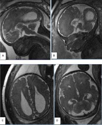
In our small series of 4000 MRI explorations in children of different ages 10 cases harboring ACC were diagnosed, accounting for 0.25%.On the other hand, in 48 pregnant women who were underwent MR investigation between 20 to 36 gestational weeks, another six cases with absence of the corpus callosum were found (Figure 1-5).
Figure 1: Axial T1mpr images show complete absence of corpus callosum and the parallel and separated bodies of lateral ventricles -giving the “ricing car appearance.
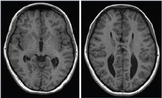
Figure 2: Coronal T2W and coronal FLAIR (tirm dark-fluid) images illustrate „ the bull’s” horns appearance .The upper corners turn up and pointed.
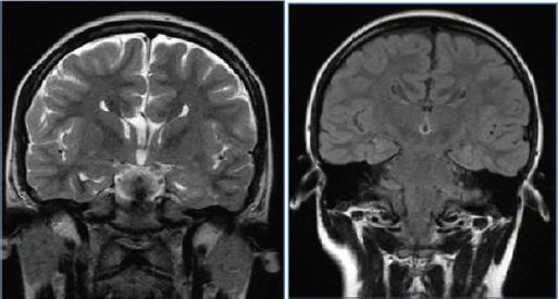
Figure 3: Sagittal (midsagittal) T1mpr shows absence of the cingulargyrus and radial pattern of the medial hemisheric sulci.
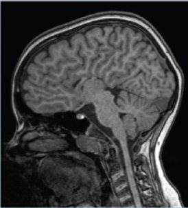
Figure 4: Coronal T2w images show big interhemispheric cyst and wide communication with enlarged and distorted third and left lateral ventricle, multiple subependymal/ subcortical heterotopias (arrow) and dysplastic cortex in both hemispheres.
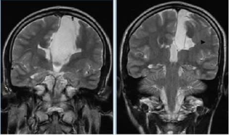
Figure 5: Fetal endocranial MR examination of fetus at 36 weeks of gestation performed in three planes (T2 trufi HQ in midsagittal, parasagittal, axial and semicoronal) shows all typical features of ACC: A) and B) absence of cingular gyrus, radial course of the medial cerebral sulci; C) prominent ventricular atria known as colpocephaly; D) frontal horns are displaced laterally giving „ the bull’s” appearance.
Conclusion
The complete corpus callosum agenesis is a frequent anomaly commonly associated with other anomalies and anatomical lesions of the brain. Genetic factors are considered to be the main cause of the malformation. There are no specific signs and symptoms of ACC. In isolated cases clinical syndromes are divers and vary in the severity of the clinical picture. Magnetic resonance imaging is the procedure of choice in the diagnosis of the disorder.
References
- Kumar P, Burton BK (2007) Congenital malformations, evidence-based evaluation and management. McGraw-Hill Professional, USA.
- Schell-Apacik CC, Wagner K, Bihler M, Wagner BE, Heinrich U, et al. (2008) Agenesis and Dysgenesis of the Corpus Callosum: Clinical, Genetic and Neuro imaging Findings in a Series of 41 Patients. Am J Med Genet A 146A(19): 2501-2511.
- Chiappedi M, Bejor M (2010) Corpus callosum agenesis and rehabilitative treatment. Ital J Pediatr 36: 64.
- Palmer EE, Mowat D (2014) Agenesis of the corpus callosum: a clinical approach to diagnosis. Am J Med Genet C Semin Med Genet 166C(2): 184-197.
- Schilmoeller G, Schilmoeller K (2000) Filling a void: Facilitating family support through networking for children with a rare disorder. Family Sci Rev 13: 224-233.
- Chiappedi M, Fresca A, Baschenis IMC (2012) Complete Corpus Callosum Agenesis: Can It Be Mild? Case Reports in Pediatrics.
- Righini A, Frassoni C, Inverardi F, Parazzini C, Mei D, et al. ( 2013) Bilateral Cavitations of Ganglionic Eminence: A Fetal MR Imaging Sign of Halted Brain Development. AJNR Am J Neuroradiol 34(9): 1841-1845.
- Neal JB, Filippi CG, Mayeux R (2015) Morphometric variability of neuroimaging features in children with agenesis of the corpus callosum. BMC Neurol 15:116.
 a Creative Commons Attribution 4.0 International License. Based on a work at www.crimsonpublishers.com.
Best viewed in


.jpg)


 Editorial Board Registrations
 Submit your Article
 Refer a Friend
 Advertise With Us
.jpg)


.jpg)


.bmp)
.jpg)
.png)
.jpg)


.png)

.png)


.png)


