- Submissions

Full Text
Orthopedic Research Online Journal
Assessment of the Possibilities of B-Mode Ultrasonography in the Diagnosis of Partial Damage to the Rotator Cuff of the Shoulder
Abdullaiev RYa* and Dudnik TA
Department of Ultrasound Diagnostics, Kharkov Medi¬cal Academy of Postgraduate Education, Ukraine
*Corresponding author: Rizvan Abdullaiev, Department of Ultrasound Diagnostics, Kharkov Medi¬cal Academy of Postgraduate Education, Ukraine
Submission: May 01, 2018;Published: August 20, 2018

ISSN: 2576-8875 Volume4 Issue2
Introduction
Diagnosis of lesions of pararticular tissue is an actual problem of traumatology and orthopedics. The shoulder joint belongs to the most vulnerable parts of the motor apparatus. Damage to the rotator cuff of the shoulder (RMP) is a complex diagnostic task, including clinical, radiological and morphological studies. Diagnosis of complete tendon ruptures does not cause difficulties in clinical examination, while partial ruptures complicate clinical evaluation. Due to the complexity of the anatomy of the rotator cuff, its ultrasound examination is a difficult task [1-3]. The method is considered an operator dependent. However, the trained specialist can achieve high acc uracy of the method [4,5].
Objective
To study the possibilities of two-dimensional echography in the diagnosis of partial damages of the rotator cuff of the shoulder joint.
Materials and Methods
Two-dimensional echography was performed in 58 patients (32 women and 35 men) with various injuries and clinical manifestations of damage to the tendons of the rotator cuff of the shoulder joint. The age of the subjects varied between 18-60 years. The comparative group consisted of 21 healthy patients without any complaints about the pathology of the shoulder joint. All patients underwent MRI and X-ray of the shoulder joint.The ultrasonic examination was carried out on Logiq 7 (QE) scanners by linear sensors with a frequency of 5-12MHz by means of polyprojection and polyposive scanning of RMP using functional ultraso nography (FUSG) and color Doppler mapping. The following ultrasonographic symptoms were assessed: the condition of the cortical layer of the head of the humerus, the thickness of the tendons of the rotator cuff of the shoulder and the tendon of the long biceps head, their structure, vascularization, integrity, the condition of the shoulder bag.The partial damage was considered with the presence of hypoechoic defects: intraarticular, extraarticular and intra-stem defects in the attachment of the tendon with a fragmentary detacment of the cartilaginous or cortical humerus.
Results
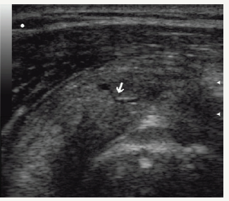
Figure 1:Partial interstitial ruptures of the rotator cuff. The line of rupture is visualized in the form of a thin hypoechogenic tubular structure (arrow).

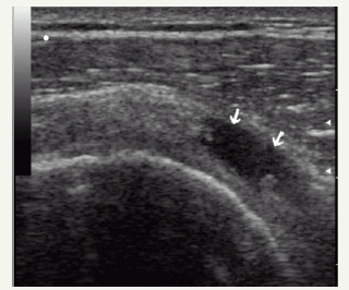
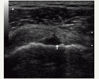
As a result ofan ultrasound study, partial damage to the tendon of the supraspinatus was diagnosed in 31 patients (53.4%), of the subspinatus - in 24 patients (41.3%), of the subcutaneous - in 8 patients (13.8%), with intra-stem injuries in 21 patients (36.2%), extraarticular lesions in 17 patients (29.3%), intraarticular lesions in 5 patients (8.6%). A partial detachment of the tendon of the supraspinatus with the bone fragment of the large tubercle from the site of its insertion was observed in 5 patients (8.6%). Partial damages of the rotator cuff of the shoulder, combined with a fracture of the large tubercle, were diagnosed in 2 patients (3.4%). Partial damages of the rotator cuff of the shoulder were accompanied by subdeltoid- subacromial bursitis in 27 (46.5%) patients (Figures 1-3). In the radiography of the shoulder joint, changes were detected in 5 patients (8.6%) with detachment of bone fragments of the large tubercle, in 2 patients (3.4%) with a fracture of the large tubercle.
The results of MRI and ultrasound did not coincide in 5.2% of cases - with synovitis of the tendon of the long biceps head. In these cases, with dynamic observation, clinical improvement coincided with the disappearance of ultrasound signs of tenosynovitis.
Figure 2:Partial interstitial ruptures of the rotator cuff is visualized in the form of a small necrogenous region with irregular, distinct contours (arrows).

Figure 3:Partial intraarticular ruptures of the rotator cuff. The rupture area faces the joint cavity (arrow). Exudation is not observed.

Conclusion
1. Ultrasonography can accurately determine the nature of damage to the elements of the rotator cuff.
2. It is an accessible and informative method for conducting dynamic observation.
Early ultrasound identification of partial damages of the rotator cuff is of great importance for determining the tactics of treatment, since it allows conservative treatment methods to be performed before the complete rupture occurs.
References
- McNally EG, Rees JL (2007) Imaging in shoulder disorders. Skeletal Radiol 36(11): 1013-1016.
- Jacobson JA (2011) Shoulder US: Anatomy, technique, and scanning pitfalls. Radiology 260(1): 6-16.
- Beggs I (2011) Shoulder ultrasound. Semin Ultrasound CT MR 32(2): 101-113.
- Smith TO, Back T, Toms AP, Hing CB (2011) Diagnostic accuracy of ultrasound for rotator cuff tears in adults: A systematic review and meta-analysis. Clin Radiol 66(11): 1036-1048.
- Abdullaev RY, Dzyak GV, Dudnik TA (2010) Ultrasonography of the shoulder joint. Kharkov, Ukraine
© 2018 Abdullaiev RYa. This is an open access article distributed under the terms of the Creative Commons Attribution License , which permits unrestricted use, distribution, and build upon your work non-commercially.
 a Creative Commons Attribution 4.0 International License. Based on a work at www.crimsonpublishers.com.
Best viewed in


.jpg)


 Editorial Board Registrations
 Submit your Article
 Refer a Friend
 Advertise With Us
.jpg)


.jpg)


.bmp)
.jpg)
.png)
.jpg)


.png)

.png)


.png)


