- Submissions

Full Text
Investigations in Gynecology Research & Womens Health
Infrared Thermography Appearance of Abdominal Wall Endometriosis: A Useful Tool for Diagnosis
Francisco das Chagas Medeiros*, Francisco Eristow Nogueira, Vietla S Rao and Dalgimar B de Menezes
Department of Gynecology, Federal University of Ceara, Brazil
*Corresponding author: FC Medeiros, Department of Gynecology, Assis Chateaubriand School Maternity, Faculty of Medicine, Federal University of Ceara, Rua Prof Costa Mendes 1608, 2nd Floor, 60416-200 Fortaleza, Ceara, Brazil, Tel: 55-85-33668524; Email: prof.fcmedeiros@ufc.br
Submission: September 09, 2017; Published: January 10, 2018

ISSN: 2577-2015Volume1 Issue4
Abstract
Case reports are significant part of the medical literature that continues to have their place in scientific journals. Recurrently they are the first evidence for new diagnosis and other medical conducts. Original and critical use of these studies can increase their values by elevating the practice of medicine. We present a new useful tool for diagnosing abdominal wall endometriosis through a teletermography in a 33 year old woman comparing with magnetic resonance and physical exam and pathological histology.
Keywords: Endometriosis; Scar endometrioma; Infrared thermography
Introduction
Endometrioma is a well circumscribed mass of endometriosis that occurs when ectopic endometrial tissue (gland and stroma), which can respond to ovarian hormonal stimulation, grows outside the uterus [1]. Endometrioma in a surgical scar develops in 0.1% of women who have undergone cesarean section, and 25% of these women have concomitant pelvic endometriosis [2]. Its clinical diagnosis in the abdominal wall has been confused with abscess, lipoma, hematoma, sebaceous cyst, suture granuloma, inguinal hernia, incisional hernia, desmoid tumor, sarcoma, lymphoma, or primary and metastatic cancer [2,3]. The diagnosis of abdominal wall endometriomas is often confused with other surgical conditions. Endometriosis is rarely seen by general surgeons and is often diagnosed on histological examination postoperatively [3]; because of the large number of potential pitfalls in its diagnosis, we present a case as seen from the perspective of a general surgeon and also by gynecologists emphasizing a new diagnosis image by skin infrared thermography. Vessels in the skin are arranged into superficial and deep horizontal plexuses and they are involved in thermoregulation, oxygen and nutritional support. The skin has a large number of functions and broad appeal spanning basic mechanistic and clinical research. Indeed, the skin can be used as a marker of normal and impaired vascular control and, owing to its accessibility and frequent involvement, is easy to investigate non-invasively. The measurement of skin temperature provides an indirect measure of blood flow and can be measured by assessing thermal convection or directly measuring skin temperature (using thermography) [4]. The technology of thermal imaging has made significant advances in recent years. It has been used in military applications widespread, but not in clinical medicine [5]. Thermography is a diagnostic method which measures temperature differences across the skin surface using a highly sensitive infrared camera. Recent development of digital infrared thermographic focal plane array cameras offers the opportunity to revitalize this diagnostic method and further improve its accuracy.
Currently, there is no consensus regarding diagnostic parameters for digital thermography in diagnosis of scar endometriosis. The aim of this paper is to examine the application of digital thermography and propose endometriosis diagnostic thermographic as compared to other modalities of exams. We report a case of scar endometriosis in a woman who underwent caesarean section diagnose by alteration in thermograph assessment.
Case Report (MIC, 33 years old)
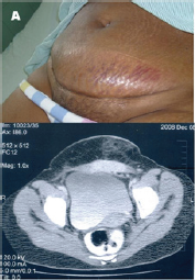
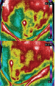
After 2 years of a cesarean section, a 33-year-old white woman presented with a 12-month history of pain in the lower right quadrant in the suprapubic region at the site of a Pfannenstiel-type laparotomy. On physical examination she had a 3-cm nodule on the cesarean scar, causing pain during menstruation. Ultrasound showed the presence of an oval-shaped hypoecogenous neoformation (Figure 1). A cytological examination indicated a cystic neo-formation removed under regional anesthesia [6]. The neoformation was firmly attached to the fascia of the rectus abdominalis muscle. Histological examination showed numerous endometriotic foci spread in an abundant fibrous tissue. They were characterized by glands in frequent cystic dilation. The glands were surrounded by a variable amount of endometrial stroma with extensive decidual changes this pattern was consistent with a secretory phase of the menstrual cycle. The patient was submitted to an infrared thermography: the woman case was kept in a warm room with temperature at 22-24 °C up to 15min. Patient complaining of a heavy pain in right-low abdomen was asked to hold up her pending abdomen for 10min. Then thermograms of the abdomino-pelvic wall were taken by using an E-25 digital infrared video camera (sensitivity at 0.2 °C; Flir Systems with data analyzing software Quick View 2) positioned at a 1.5m far from the pelvis which provides the thermo pictures and reads the temperatures without touching the subject. All the data were collected in a computer. A hot area (Ar1 - white colored with black dots) showing temperatures from 34.0 °C to 35.4 °C, was spotted on the skin over the right inguinal ligament, and considered hyperthermic, when compared to the same opposite region, used as control, showing temperatures from 32.4 °C-33.7 °C (Figure 2). Thermographic studies use the ipsilateral parts of the body scanning as self-control, as well as the sudden variations of temperature (±0.6 °C) to determine hyperthermic or hypothermic areas. These temperatures gradients are out of our skin heat perception and Thermographic scanning made it possible to find the hyperthermia area.
Figure 1: Panel A.

Figure 2: Panel B.

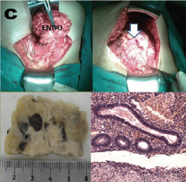
The surgical approach was through surgical incision and exeresis of the endometrioma (Figure 3). At the Panel 3 one may see the macroscopic and microscopic diagnosis of endometriosis by findings by viewing endometrial glands and stroma.
Figure 3: Panel C.

Discussion and conclusion
The imaging modalities are non-specific but useful in determining the extent of the disease and planning of operative resection, especially in recurrent and large endometriosis lesions suspicion. Compared with ultra-sonography, the theletermography is less invasive; it needs no physical contact between the probe and the skin of the patient, thus avoiding discomfort, possible physical trauma, and psychological embarrassment. It has the advantage of detecting minimal nodular alterations. Our experience in selected cases leads us to believe that theletermogrphy was as a good method to the diagnosis of varicocele as well as to endometriosis, especially for those patients who are mildly symptomatic or who have no symptoms [7].
Referencess
- Koger KE, Shatney CH, Hodge K, McClenathan JH (1993) Surgical scar endometrioma. Surg Gynecol Obstet 177(3): 243-246.
- Singh KK, Lessells AM, Adam DJ, Jordan C, Miles WF, et al. (1995) Presentation of endometriosis to general surgeons: a 10-year experience. Br J Surg 82(10): 1349-1351.
- Seydel AS, Sickel JZ, Warner ED, Sax HC (1996) Extraplevic endometriosis: diagnosis and treatment. Am J Surg 171(2): 239-241.
- Wright CI, Kroner CI, Draijer R (2006) Non-invasive methods and stimuli for evaluating the skin's microcirculation. J Pharmacol Toxicol Methods 54(1): 1-25.
- Izhar, LilaI Z, Petrou, Maria (2012) Thermal imaging in medicine in advances in imaging and electron physics. 171: 41-114.
- Medeiros FC, Cavalcante DI, Medeiros MA, Eleuterio J (2011) Fine needle aspiration cytology of scar endometriosis: Study of seven cases and literature review. Diagn Cytopathol 39(1): 18-21.
- Nogueira FE, Medeiros FD, Barroso LV, Miranda EP, de Castro JD, et al. (2009) Infrared digital telethermography: a new method for early detection of varicocele. Fertil Steril 92(1): 361-362.
© 2018 Francisco das Chagas Medeiros, et al. This is an open access article distributed under the terms of the Creative Commons Attribution License , which permits unrestricted use, distribution, and build upon your work non-commercially.
 a Creative Commons Attribution 4.0 International License. Based on a work at www.crimsonpublishers.com.
Best viewed in


.jpg)


 Editorial Board Registrations
 Submit your Article
 Refer a Friend
 Advertise With Us
.jpg)


.jpg)


.bmp)
.jpg)
.png)
.jpg)


.png)

.png)


.png)


