- Submissions

Full Text
Experimental Techniques in Urology & Nephrology
FlexDex™: A Novel Articulated Laparoscopic Instrument to Perform Renorrhaphy
Hari Vigneswaran* and Simone Crivellaro
Depar™ent of Urology, University of Illinois at Chicago, USA
*Corresponding author: Hari Vigneswaran, Depar™ent of Urology, University of Illinois at Chicago, USA
Submission: September 13, 2017; Published: November 13, 2017

ISSN: 2578-0395 Volume1 Issue2
Abstract
This is a case report series of two patients undergoing laparoscopic partial nephrectomy using a novel laparoscopic instrument with increased degrees of freedom for more optimal needle control. The laparoscopic Flexdex™ articulating needle driver is the first ever documented partial nephrectomy using this instrument which should improved ergonomics during renorrhaphy and is a potential tool to increase laparoscopic comfort and ease with surgeons.
Abbreviations: MIS: Minimally Invasive Surgical; GFR: Glomerular Filtration Rate; NSS: Nephron Sparing Surgery
Background
Surgery in the 21st century relies primarily on minimally invasive surgical (MIS) techniques thanks to the rapid evolution in technology and innovation. In terms of broad classification, there exist two spectra of MIS. The traditional laparoscopic MIS first described in 1985, which uses both optical and haptic feedback, with hand-held, light weight, cost-effective mechanical instruments that have been described to have steep learning curves due to the limited four degrees of motion. Conversely, the other end of the spectra of MIS uses the expensive robotic assisted telemanipulating surgical instruments, which rely on visual cue and are designed intuitively to follow the natural seven degrees of hand and wrist motion that can come close to mimicking open surgery.
Prior to the popularization of the robot a majority of the MIS partial nephrectomies were done with traditional laparoscopy, however as the urologist became comfortable with robotic surgery, robotic assisted partial nephrectomies began to comprise the majority of MIS trea™ent [1]. Literature supports that the learning curve may not be as steep with robotic surgery [2] as well as the benefit of improved perioperative outcomes including complete resection of margins, shorter warm ischemia time, and less conversion to open surgery [3]. The biggest drawback of robotic assisted partial nephrectomy compared with traditional laparoscopy is cost [4].
The FlexDex™ Surgical laparoscopic needle driver, an instrument designed to model the seven degrees of motion the robotic arms mimics while also maintaining traditional laparoscopic advantages may propose an improved solution to the trea™ent of partial nephrectomy. The following cases aim to demonstrate the implementation feasibility of this instrument in clinical practice.
Case Presentation
This is a report of two laparoscopic partial nephrectomy cases done with the articulating FlexDex™ laparoscopic needle driver. In terms of published literature, these are the first ever performed partial nephrectomies utilizing these new articulating laparoscopic instruments. Of note both of these patients involved in these cases presented to a community hospital that did not have the infrastructure for the da Vinci Surgical System or any other robotic surgical system. The surgeon conducting these cases had extensive experience in traditional laparoscopy and was very comfortable performing laparoscopic partial nephrectomies.
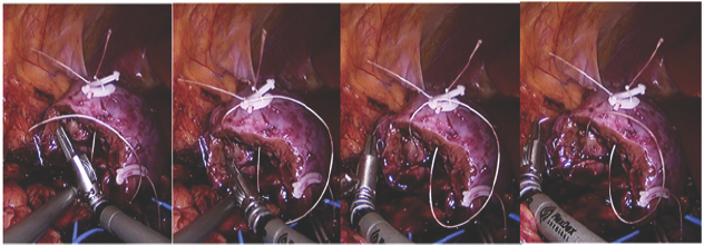
It is the opinion of these authors that the primary advantage during a robotic assisted laparoscopic partial nephrectomy compared to pure laparoscopic surgery is most clearly elucidated during the renorrhaphy. The proposed advantage is found with the additional degrees of motion that the wrist provides with the robot allowing for a faster, more precise reconstruction of the renal parenchyma. For the presented cases, the Flexdex™ laparoscopic needle driver was used during this portion of the operation (Figure 1).
The first patient is a 56-year-old-male with a significant medical history for HIV, hypertension and multiple decade history of smoking. He was referred to the urology clinic after one episode of gross hematuria. At presentation he denied any other urinary symptoms, flank mass or pain. He underwent a CT scan which revealed a 4.7cm right lower pole complex cystic mass with septations, solid components abutting the collecting system and approximately 25 Hounsfield units of enhancement with intravenous contrast. These findings were highly concerning for malignancy and after a collaborative discussion with the surgical team and the patient a minimally invasive nephron sparing was planned.
Figure 1: Laparoscopic renorrhaphy with the FlexDex™ articulating needle driver to sew using a running vicryl stitch with Weck Hem-o-lok clip on either end of reconstruction. From case #2

Our second case describes a pleasant 65-year-old female who has a significant medical history for osteoarthritis, hypertension, and a total abdominal hysterectomy for benign disease. She presented to the urology clinic with gross hematuria. Her history was significant for a past of smoking. On physical she denied any abdominal pain or palpable mass. She underwent a hematuria work up with a CT scan, which revealed an endophytic, 2.7 by 2.9 by 4.8cm right lower pole mass that was near the lower pole collecting system. Radiographic findings were highly suspicious for renal cell carcinoma. Similarly, a collaborative discussion took place with the patient and surgical team evaluating the trea™ent options and the decision was made to proceed with a minimally invasive nephron sparing surgery.
Given the similar location of the tumors in the right lower pole of the kidney for these two cases, the majority of the steps were nearly identical, however after gaining intra operative experience with the Flexdex™ the second case benefited primarily in optimal port placement. In both cases the patient was positioned in the left lateral kidney position on the operating table. Ports included two 12mm ports and two 5mm ports. The major difference between the subsequent case was understanding the need for port placement closer to the kidney to optimize the fulcrum needed when using the Flexdex™ needle driver. Colon mobilization and hilar dissection took place using standard non articulating laparoscopic instruments. Once hilar control was established Gerota's Fascia was exposed and the respective kidney tumor sites were delivered into the field of vision.
Once all equipment was prepared, including the wrist attachment for the Flexdex™ instrument already applied to the operating surgeon, the vascular elements were then clamped. Subsequently a cold knife excision of the tumors took place. Once the tumor bed was exposed the reconstruction took place using a 15cm 3.0 vicryl suture for the medullar and multiple 15cm 2.0 vycrylsutures, both with Weck Hem-o-lok clips sliding technique as normally performed during a robotic partial nephrectomy. Once this renorrhaphy was complete the vessels were unclamped and the repair was examined for bleeding. In both surgeries hemostasis was obtained, however clamping times differed with the first case taking approximately 40 minutes and the second case taking 23 minutes.
In the first case blood loss was 200ml as compared to 10ml for the second case. Pathology for both cases revealed clear cell carcinoma. Post operative creatinine and estimated glomerular filtration rate (eGFR) on day of discharge was equivalent to preoperative levels for both patients. Both patients required approximately two post operative days to recover in the hospital and both were without complications or transfusions as defined in the literature [5].
Discussion
These two cases demonstrated the feasibility of performing laparoscopic partial nephrectomy using the novel articulating needle driver. After just one use, it was clear this instrument is different from typical laparoscopic instruments in that there needs more working room to allow for fulcrum optimization of the additional degrees of motion preserved within the wrist. However, it is evident that with the first few uses an experienced laparoscopist can implement this tool, which can be especially useful with laparoscopic suturing. Immediate differences were noted in the ability to control needle advancement and maintain a rhythm similar in form to robotic suturing.
Discussing the different modalities for MIS in regards to partial nephrectomy provides an excellent platform to study the advantages of instruments like the Flexdex™. Although there is data to support robotic superiority to laparoscopic partial nephrectomy in regards to shorter warm ischemia times, lower conversion to open, and improved resection margins, there is also data which shows no difference [3,6]. Proponents of laparoscopy show that in the right hands laparoscopic surgery can lead to shorter warm ischemia times, lower rates of conversion to open, smaller change of eGFR, and shorter length of stay [7].
Despite this controversy it has been suggested that since the utilization of the robot there has been an increase in the utilization of nephron sparing surgery (NSS), the appropriate management for T1 tumors [8]. Although studies vary, it is well established that the learning curve for laparoscopy is higher than for robotic surgery and instruments like the Flexdex™ may help to bridge this difference. Giving providers a chance to utilize a more intuitive laparoscopic instrument may ensure a higher level of comfort and the ability to provide MIS NSS, the standard of care for these lesions.
Conclusion
This study showed that the laparoscopic articulating needle driver was safe and improved laparoscopic suturing efficiency. Although this instrument will need to be studied in larger trials, preliminary evidence suggests that this instrument may improve MIS methods. The instrument may provide an alternative to the surgeon that is both more intuitive to use than traditional laparoscopy and more cost effective than robotic surgery.
References
- Ghani KR, Sukumar S, Sammon JD, Rogers CG, Trinh QD, et al. (2014) Practice patterns and outcomes of open and minimally invasive partial nephrectomy since the introduction of robotic partial nephrectomy: results from the nationwide inpatient sample. J Urol 191(4): 907-912.
- Hanzly M, Frederick A, Creighton T, Atwood K, Mehedint D, et al. (2015) Learning curves for robot-assisted and laparoscopic partial nephrectomy. J Endourol 29(3): 297-303.
- Leow JJ, Heah NH, Chang SL, Chong YL, Png KS, et al. (2016) Outcomes of Robotic versus Laparoscopic Partial Nephrectomy: an Updated MetaAnalysis of 4,919 Patients. J Urol 196(5): 1371-1377.
- Hyams E, Pierorazio P, Mullins JK, Ward M, Allaf M, et al. (2012) A comparative cost analysis of robot-assisted versus traditional laparoscopic partial nephrectomy. J Endourol 26(7): 843-847.
- Pareek G, Hedican SP, Gee JR, Bruskewitz RC, Nakada SY (2006) Metaanalysis of the complications of laparoscopic renal surgery: comparison of procedures and techniques. J Urol 175(4): 1208-1213.
- Ng AM, Shah PH, Kavoussi LR (2017) Laparoscopic Partial Nephrectomy: A Narrative Review and Comparison with Open and Robotic Partial Nephrectomy. J Endourol doi: 10.1089/end.2017.0063.
- Choi JE, You JH, Kim DK, Rha KH, Lee SH (2015) Comparison of perioperative outcomes between robotic and laparoscopic partial nephrectomy: a systematic review and meta-analysis. Eur Urol 67(5): 891-901.
- Maurice MJ, Zhu H, Kim SP, Abouassaly R (2016) Increased use of partial nephrectomy to treat high-risk disease. BJU Int 117(6B): 75-86.
© 2017 Hari Vigneswaran, et al. This is an open access article distributed under the terms of the Creative Commons Attribution License , which permits unrestricted use, distribution, and build upon your work non-commercially.


.jpg)


 Editorial Board Registrations
 Submit your Article
 Refer a Friend
 Advertise With Us
.jpg)


.jpg)


.bmp)
.jpg)
.png)
.jpg)


.png)

.png)


.png)


