- Submissions

Full Text
Experiments in Rhinology & Otolaryngology
A Modified Technique of Rhinoplasty Using Cortical Bone Graft to Correct Saddle Nose Deformity with Loss of Septal Cartilage
Manu Malhotra1*, Saurabh Varshney2, Poonam Joshi2, Shubhankur Gupta2, Rashmi Malhotra3 and Vivek Singh4
1 Department of Otolaryngology AIIMS, India
2 Department of Otolaryngology & HNS (ENT), AIIMS, India
3 Department of Anatomy, AIIMS, India
4 Department of Orthopedics, AIIMS, India
*Corresponding author: Manu Malhotra, Associate Professor, Department of Otolaryngology AIIMS, Veerbhadra Marg, Rishikesh, Uttarakhand-240201, India
Submission: May 8, 2018;Published: July 25, 2018

ISSN 2637-7780 Volume2 Issue1
Abstract
Several types of graft materials are used in correction of correction of saddle nose deformities but autologous tissues including remain the materials of choice due to ease of availability and biocompatibility. This article describes a case of saddle nose deformity with loss of Septal cartilage due to septal abscess which was corrected by a modified technique of augmentation Rhinoplasty using tri-cortical iliac crest graft for dorsum, combined with monocortical iliac graft to form a ‘Golf stick’ for Septal augmentation and, modified Goldman’s tip-plasty for tip correction.
Keywords: Rhinoplasty; Osteotomy; Graft; Autograft
Introduction
Nose is the most prominent structure on the face which makes it amenable to trauma consequently the nasal bone is the most commonly fractured bone in the human skeleton and most cases of septal hematomas and abscesses arise from the nasal injury [1]. The cartilaginous septum and maxillary bone crest form the main support of the lower two thirds of the nasal dorsum. If there is insufficient septalcartilage to give support, nasalsaddling of various degrees will result. Thisis commonly seen after septal haematoma,septal surgery or trauma,when haematoma is infectedleading to formation of abscess, necrosis of septal cartilage and subsequent nasal collapse. Immediate grafting is advocated by somebut in most instances grafting ofthe dorsum is deferred until the degree of saddling is evident [2]. There are various grafts and implants that can be used to restore the volume and structural integrity of the nose.
The available biomaterials can be divided into three categories:
A. Autografts- which arederived from the patient and include cartilage,bone, fascia and dermis
B. Homografts– Which are derived from tissues donated by members ofthe same species and includes irradiated cartilage and acellular dermis.
C. Alloplastic implants - that are synthetic implants (biocompatible polymers) with a variety of applications in plastic surgery.
[3] Due to excellent bio-compatibility, the ability to reconstruct like tissue with like tissue, and the relatively low risk profile, autologous grafts are usually preferred when such material is available insufficient quantity to achieve adequate augmentation [4]. Iliac crest bone graft has the advantage that it is available in bulk amountand is very useful in case of severe saddle deformity [2]. We hereby present a case of post septal abscess saddle nose deformity with tip depression, columellar retraction and necrosed septal cartilage leading to collapsed nasal valve with nasal obstruction. Augmentation septo-rhinoplasty was done using tri-cortical iliac crest graft combined with mono-cortical iliac graft to form a ‘Golf stick’. The case is unique because of the different technique adopted to resolve a difficult clinical and aesthetic situation.
Case Report
An 18 years old male patient presented with the complaints of nasal obstruction and saddle nose deformity since last 2 years. He gave history of blunt trauma of nose 2 years back which was followed by the swelling of dorsum and nasal obstruction. He took medical consultation (records of which are not available) a few weeks later, when his obstruction was not relived. There is history of incision and drainage of pus from nasal septum (records not available).Following which his nasal bridge got depressed. On examination the dorsum of the nose was saddled, tip bullous, and the septum was thickened bilaterally on anterior rhinoscopy (Figure 1). Air entry to nose was decreased due to collapse of valve on both sides.A computerised tomographic scan was performed with 3D reconstruction, which confirmed septal thickening and depression of bony and cartilaginous vaults (Figure 2). Rest of the ear, nose, throat and head-neck, along with systemic examination was normal. The patient consented for iliac crest graft after all the possible sites of autologous grafts and possibility of alloplastic grafts along with their advantages and disadvantages were explained to him.
Figure 1:

Figure 2:

Figure 3:

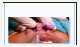
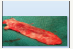
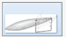
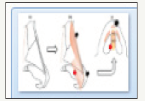
External septo Rhinoplasty was planned under general anaesthesia. Exposure was through ‘V’ shaped mid-columella incision continued with a marginal incision (Figure 3). The skin of the dorsum was elevated using sharp dissection with fine scissors. Medial, lateral and horizontal osteotomies were performed using 3mm osteotome so as to correct flaring of dorsum after correction(Figure 4). Upper lateral cartilages and lower lateral cartilages were separated in midline to accommodate the septal graft. A bi-cortical iliac crest was harvested for correction of saddling and a 2cm X 1.5cm straight chip of mono-cortical bone was harvested from the same site.The crest graft was measured for the size of correction and drilled into the shape of an inverted boat (Figure 5). The Mono-cortical graft was cut to the appropriate size and fixed to the dorsal graft. Upper and lower lateral cartilages were separated in midline and space between septal perichondrium was opened. The graft assembly was installed with flat mono-cortical graft in space between septal muco-perichondrium on both sides as shown in (Figure 6). The assembly was secured with absorbable sutures. A modified Goldman’s tip-plasty was further performed to sharpen the tip. The pre and post-operative pictures are shown in (Figure 1). When examined after two, and then four months there was no edema over face, skin over the graft was normal and the airway had become patent. The pain at donor site had subsided by the end of a month and patient was satisfied with surgical outcome.
Figure 4:

Figure 5:

Figure 6:

Discussion
Autologus grafts for augmentation Rhinoplasty include cartilage, bone, fascia and dermis. They induce no immune response and are therefore biocompatible. Although cartilage grafts have been first material of choice, they are useful mostly in mild deformities and sometimes may cause post-operative defects.The most frequent defects are graft displacement and asymmetry,tendency to curl (especially rib cartilage), a visible step, and under or overcorrection. Alloplastic implants are also used for nasal augmentation. Their advantages are ready availability in shapes and sizes and no donor site morbidity. These materials have a high rate of complications like extrusion, malposition, or unnatural appearance. Autologus bone grafts are the best option for reconstruction of wide defects of the nose, maintaining the tip projection with minimal airway problems [5].
Bone graft provides a durable and safe reconstruction.Tissue for augmentation Rhinoplasty has been obtained from cranial bone, ribs, iliac bone and olecranon. Calvariasbone is the most commonly used bone graft. It has numerous advantages as a donor site: itresorbs less, is an easy to hide donor site, and does not bend. However, serious complications like intracerebral haematoma, superior sagittal sinus laceration have been reported [5-7]. Iliac crest bone graft has the advantage that it is available in bulk amount and is very useful in case of severe saddle deformity involving both the cartilaginous and bony dorsum. The graft is very stable over time, it does not change shape or bend like cartilage, can be easily harvested, incision is hidden, and the donor and recipient sites are different. The disadvantages with this graft are that the reshaping is difficult; due to hard nature of bone and the most significant morbidity is pain. Bone grafting from the iliac crest is a relatively benign procedure in terms of patient satisfaction [2]. Though some have questioned the long term survival of iliac crest graft, but Karakaoglan and Usual reviewed 14 patients who underwent iliac crest bone grafts to augment the nasal dorsum, and with an average follow up of one to four years, reported no significant resorption [6]. Several other authors have in past recommended iliac crest graft on basis of good success rates [6-8].
The case reported not only had a gross saddle nose deformity but collapse of nasal valve leading to serious obstruction. The reconstruction technique adopted was unique because it used iliac crest graft fixed in a new fashion with a plate of mono-cortical graft along with a modified Goldman’s tip-plasty.
Conclusion
The autologous iliac crest bone graft gives an excellent outcome in augmentation Rhinoplasty with minimum complications and it is highly suitable for that case in which there is severe degree of nasal dorsum saddling. The modified technique of augmentation Rhinoplasty, tip-plasty and Septal augmentation using golf stick shaped assembly is can be useful for restoration of normal airway and aesthetics of face.
References
- Nwosu JN, Nnadede PC (2015) Nasal septal hematoma/abscess: management and outcome in a tertiary hospital of a developing country. Patient Preference and Adherence 9: 1017-1021.
- Saeed M, Hussain Z, Mian FA (2012) Augmentation rhinoplasty with autologus iliac crest bone graft. APMC 6(1): 18-21.
- Ali Sajjadian A, Rubinstein R, Naghshineh N (2010) Current Status of grafts and implants in rhinoplasty: part I. autologous grafts. Plast Reconstr Surg 125(2): 49e-40e.
- Dresner HS, Hilger PA (2008) An Overview of nasal dorsal augmentation. Semin Plast Surg 22(2): 65-73.
- Chauhan DS, Guruprasad Y (2012) Cortical tibial bone graft for nasal augmentation. J Maxillofac Oral Surg 11(2): 186-190.
- Karakaoglan N, Usyal OA (1998) Use of iliac bone graft for saddle nose deformity. Auris Nasus Larynx 25(1): 49-57.
- Sarukawa S, Sugawara Y, Harii K (2004) Cephalometric long term followup of nasal augmentation using iliac bone graft. J Craniomaxillofac Surg 32(4): 233-235.
- Goodman WS, Gilbert RW (1985) Augmentation rhinoplasty a personal view. J Otolaryngol 14(2): 107-112.
© 2018 Manu Malhotra. This is an open access article distributed under the terms of the Creative Commons Attribution License , which permits unrestricted use, distribution, and build upon your work non-commercially.
 a Creative Commons Attribution 4.0 International License. Based on a work at www.crimsonpublishers.com.
Best viewed in


.jpg)


 Editorial Board Registrations
 Submit your Article
 Refer a Friend
 Advertise With Us
.jpg)


.jpg)


.bmp)
.jpg)
.png)
.jpg)


.png)

.png)


.png)


