- Submissions

Full Text
COJ Nursing & Healthcare
A Study on the Relationship between Iron Reserves and the Intensity of Coronary Artery Stenosisin Angiography Candidates
Faride Rashidi1, Khalil Mahmoodi2* and Mehran Tahrekhani3
Tabriz University of Medical Sciences, Iran
Department of Internal Medicine, Zanjan University of Medical Sciences, Iran
Department of Nursing, Zanjan University of Medical Sciences, Internal
*Corresponding author: Khalil Mahmoodi, Associate Professor, Department of Internal Medicine, School of Medicine, Zanjan University of Medical Sciences, Zanjan, Iran, Tel: 09122410801; Email: kimahmood@gmail.com
Submission: February 22, 2018;Published: March 22, 2018

ISSN: 2577-2007Volume2 Issue1
Abstract
Various risk factors including blood iron may create coronary artery diseases and lead to myocardial infarction. There are controversies with regard to the impact of blood iron on myocardial infarction. Therefore, the aim of this paper was to investigate the relationship between iron reserves and the intensity of coronary artery stenos is among angiographic candidates in Zanjan, Iran. This was a cross sectional study. Samples were consisted of patients who were hospitalized for diagnostic coronary angiography in hospitals in an urban area of Iran. A convenient sampling method was used to recruit samples via interviews and laboratory examinations for FBS, iron, TIBC, ferritin, creatinine serum, CBC, cholesterol, HDL and LDL. The samples were divided into control and intervention groups. After coronary angiography, the intervention group was evaluated by four different methods including the extent score, stenos is score, vessel score and Duke CAN Index. The samples were consisted of 89 men (60.1%) and 59 women (39.9%). The levels of ferritin (p=0.003) and iron (p=0.002), and transferrin saturation percent (p=0.002) showed significant differences between males and females (p=0.004).
The linear relationship between ferritin and the intensity of coronary artery stenosis was obtained by the first method (p=0.006), second method (p=0.002), third method (p=0.001) and forth method (p=0.003). No statistically significant relationships were reported between the transferrin saturation percent and the intensity of stenosis by the above-mentioned methods (p=0.32, p=0.89, p=0.77 and p=0.79). This study showed that the intensity of coronary artery stenosis had a relationship with the body iron reserves. This relationship only was available for ferritin and not for relationships between iron and the transferrin saturation percent. Therefore, ferritin could be a more reliable index for predicting the intensity of coronary artery stenosis.
Keywords: Iron reserves; Coronary artery; Angiography; Ferritin serum
Background
Coronary artery disease (CAD) is the most rampant disease that leads to mortality and morbidity. It leads to numerous economic losses and has many effects on individuals' quality of life [1]. CAD begins at the early stages of life, but its manifestations are revealed after a long period of incubation. Environmental, behavioral, genetic and dietary factors are some risk factors for CAD [2]. The prevention of CAD and reduction of the patients' mortality rate are the concerns of the healthcare system across the world. Achieving such a goal requires improving our understanding of risk factors related to them [3-5]. Coronary risk factors are consisted of hereditary and nonadjustable ones such as age, gender, family background and adjustable ones including obesity, smoking, blood pressure and diabetes mellitus [6]. Another risk factor for CAD is the body iron reserves [7]. Many studies focused on the direct and indirect role of the body iron reserves in the creation and progression of CAD. Paradoxical results have been found on the role of the body iron reserves on CAD [8-11]. Sullivan [12] for the first time proposed that the body iron reserves can probably have a positive relationship with CAD [12].
The assessment of the plasma ferritin level is a suitable diagnostic method for evaluating iron balance in the body [13,14]. Since serum ferritin is an iron storage protein and one of most powerful factors in creation and progression of carotid artery diseases, its relationship with CAD has not been clearly understood. In a study by Khadem Vatan and Ahmad Pour (2015) only the level of coronary artery stenosis and the amount of ferritin serum were investigated [3]. Therefore, in this study, four various methods were applied for the assessment of iron storage protein (ferritin serum) and its relationship with CAD among patients admitted for coronary angiography to Zanjan hospitals.
Method
This was a cross sectional study conducted with 148 patients undergoing diagnostic coronary angiography. This study was authorized by the research council affiliated with Zanjan University of Medical Sciences Samples were all patients who were hospitalized for diagnostic coronary angiography in Zanjan hospitals in 2012. A convenient sampling method was used to recruit the patients. Patients who had myocardial infarction during the last 3 months, being diagnosed with diabetes, being caught by infectious diseases in the week before hospitalization and had gastrointestinal bleeding in the week before hospitalization were excluded from the study. Eligible patients were informed of the study's aim and process. If they were willing to take part in this study, signed written informed consent form. Data gathering was conducted using face to face interviews with the patients for health-related and demographic information, data collected after watching the patients' coronary angiography movies and performing laboratory examinations. The demographic and health-related data was age, gender, height, weight and the body mass index (BMI), Cholosterol, LDL, HDL and TG levels, the family background of CAD, blood pressure and smoking habits.
The patients' blood samples were taken after 12 hours of fasting and were sent to the laboratory before coronary angiography. The blood samples were used for assessing the patients FBS, iron, TIBC, ferritin, creatinine, CBC, cholesterol, HDL, LDL and TG. After coronary angiography, the movies of the procedure were stored in CDs and kept for further reviews. The movies were reviewed by a cardiologist and the patients were divided into control and intervention groups. The intervention group had a remarkable blockage (higher than 50%) in at least one of coronary arteries, but the control group showed no remarkable stenosis (lower than 50%). The patients were scored by using four methods and received scores for each method. The scoring process in each method was defined as follows:
A. The first method (Vessel score)-The numbers of arteries with remarkable stenosis (higher than 50%) were determined and each artery with remarkable stenosis received a score between 0 to 3.
B. The second method (Stenosis score)-All eight coronary arteries were scored in terms of the intensity of the stenosis as follow: stenosis between 1%-49%=score 1, 50%-74%=score 2, 75%-99%=score 3 and for complete stenosis=score 4. The sum of the scores was calculated and the maximum score for a patient with eight complete blocked arteries were 32.
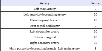
C. The third method (Extent score)-It was one of the most accurate scoring methods for checking the intensity of stenosis, the percent of the arterioscoloris plaque as the length and stenosis in each coronary artery was multiplied by a certain coefficient defined separately for each coronary artery. Coefficients for each of the main arteries were shown in Table 1.
Table 1: The coefficients for main coronary arteries.

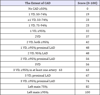
D. The fourth method (Duke CAD Index)-Given the number of arteries with remarkable stenosis (higher than 50%) as well as the anatomical place of the stenosis in terms of being proximal or distal, scores were determined (Table 2).
Table 2: Duke scoring method.

After the completion of the data collection process, data was entered to the SPSS statistical software v.18. The relationship between qualitative variables was assessed using the chi-square test. Also, the difference between groups' mean was assessed using the student t-test. Moreover, confounding variables such as age, gender, BMI, smoking, hypertension, lipid profile and the Glomeruli Filtration Rate (GFR) were analyzed using the linear regression test. P<0.05 was considered statistically significant.
Results
In this study 148 patients were recruited as 89 patients (60.1%) were men and 59 patients were women (39.9%). Their mean age was 57 years with a range of 30-77 years. (Table 1-4). The linear relationship between ferritin and the intensity of coronary artery stenosis was obtained by the first method (p=0.006), second method (p=0.002), third method (p=0.001) and forth method (p=0.003) (Table 3). However, no statistically significant relationship was reported between the iron level and the intensity of coronary artery stenos is in the first method (p=27.0), second method (p=42.0), third method (p=0.37) and fourth method (p=13.0) and also between the transferrin saturation percent and the intensity of stenosis in the first method (p=79.0), second method (p=77.0), third method (p=0.89) and the fourth method (p=32.0) (Table 4).
Table 3: The relationship between the intensity of coronary artery stenosis and iron (ferritin) reserves.

Table 4: The relationship between the intensity of coronary artery stenosis and iron reserves as the transferrin saturation percent.

The levels of ferritin (p=0.003) and iron (p=0.002), and transferrin saturation percent (p=0.002) showed significant differences between males and females. Linear relationships between other risk factors for CAD and the intensity of stenos is were also calculated in which cholesterol and the intensity of stenosis had a meaningful relationship in terms of stenosis (p=04.0), vessel (p= 04.0) and extent methods (p= 0.013). On the other hand, no statistically meaningful relationships were reported between age, BMI, GFR and the intensity of stenosis (p<0.05). Differences between the intensity of stenosis in patients with and without hypertension were calculated in four methods as follow: stenosis (p=0.047), vessel (p=0.05) and extent methods (p=0.039), but they were not meaningful in the Duke scoring method (p<0.05). The difference between the intensity of stenosis in smoker patients and patients with family history of CAD was not statistically significant. Moreover, the linear relationship between the number of white blood cells and the intensity of coronary artery stenosis was statistically significant in terms of stenosis (p=0.036) and extent methods (p=0.037). Confounding factors such as hypertension, smoking and family backgrounds were assessed using the linear regression method in which the relationship between ferritin and the intensity of coronary artery stenosis was statistically significant via the four methods (p=0.003, p=0.002, p=0.002 and p=0.004).
Discussion
This study showed that the intensity of coronary artery stenosis had a relationship with the body iron reserves. This relationship was correct only for ferritin and the intensity of stenosis. No statistically significant relationship was reported between iron and transferrin saturation percent. With regard to the role of Iron in ischemic diseases, controversial results are available. In the study by Bagheri et al. [1] (2013) in Sari, the level of iron in the blood serum of patients with arterioscoloris was high. The amount of iron had a direct relationship with the intensity of CAD, which was in line with the findings of our study. The findings of our study were also confirmed by those of other studies [1,10,15]. Haj Ahmadi Pour & khadem Vatan [3] showed no statistically significant relationship between the coronary artery stenosis and ferritin level in males and females. Also, no statistically significant relationship was reported between the involvement of the coronary arteries and iron serum, which probably could be due to the application of a different method for data collection [3].
The study conducted by Heidari showed that the comparison between iron reserves in males and females had a significant difference. This can be a good example for gender differences and its impact on the occurrence of ischemic diseases. Heidari reported a statistically significant relationship between the ferritin level and CAD in male patients but no statistically significant relationship could be detected among females [16]. The findings related to male patients were similar to the findings of this study. Ali Pour Moghaddas [13] a statistically significant relationship was observed between ferritin serum and CAD in male patients [13]. Through investigating the blood iron concentration, some insight could be gained into the intensity of coronary artery thrombosis. Also based on the rates of laboratory (clinical) subsets which are used to determine the blood iron concentrations, such as, ferritin serum, the patients with coronary artery thrombosis could be screened and, in turn, treated effectively. Given the controversial results of different studies, the relationship between iron reserves and the intensity of coronary artery stenosis needs further studies with higher sample sizes for removing confounding factors influencing both these variables.
Conclusion
According to this study, the intensity of coronary artery stenosis was related to the body iron reserves. This relationship was only correct for ferritin and the intensity of stenosis and iron and the transferrin saturation percent did not did not show significant relationships. Therefore, ferritin is a reliable index for predicting the intensity of coronary artery stenosis. Given the fact that the body iron reserves play as the risk factor for the creation of CAD, its amount in men's blood should be evaluated and appropriate interventions such as phlebotomy should be conducted [17]. The level of ferritin serum in men is reduced to half by one unit blood donation a year [18,19].
Limitations
Given the fact that iron and ferritin are present in the acute phase of the disease, the CRP level could be measured for tracking probable infections. Moreover, small sample size and the evaluation for the intensity of stenosis based on the subjective perspective of evaluator were other limitations of this study.
The most accurate method for assessing the iron levels is the measurement of the ratio between transferrin to ferritin receivers. However, due to the economic limitations and high price of the measurement kits, the measurement of this index was impossible.
Acknowledgment
We would like to thank all patients for their participation in this study. Our sincere gratitude belongs to the deputy of research affiliated with Zanjan University of Medical Sciences for financial support to this research project.
References
- Bagheri B, Shokrzadeh M, Mokhberi V, Azizi S, Khalilian A, et al. (2013) Association between Serum Iron and the Severity of Coronary Artery Disease. Int Cardiovasc Res J 7(3): 95-98.
- Tsimikas S, Hall J (2012) Lipoprotein (a) as a potential causal genetic risk factor of cardiovascular disease: a rationale for increased efforts to understand its pathophysiology and develop targeted therapies. J Am Coll Cardiol 60(8): 716-721.
- Haj ahmadipoor Rafsanjani M, Khadem vatani K (2015) Survey of the iron store with coronary artery disease in patients undergoing selective coronary angiography. Uromiye University of Medical Sciences 9(26): 764-774.
- Muñoz Bravo C, Gutiérrez Bedmar M, Gómez Aracena J, García Rodríguez A, Fernández-Crehuet Navajas J (2013) Iron: Protector or Risk Factor for Cardiovascular Disease? Still Controversial. Nutrients 5(7): 2384-2404.
- Bozzini C, Girelli D, Tinazzi E, Olivieri O, Stranieri C, et al. (2002) Biochemical and Genetic Markers of Iron Status and the Risk of Coronary Artery Disease: An Angiography-based Study. Clin Chem 48(4): 622-628.
- Sonia SA, Shofiqul I, Annika R, Maria GF, Krisela S, et al. (2008) Risk factors for myocardial infarction in women and men: insights from the INTERHEART study. Eur Heart J 29(7): 932-940.
- Beata P, Tomasz S, Bartiomiej P, Martyna O, Slawomir P, et al. (2013) Iron status and survival in diabetic patientswith coronary artery disease. Diabetes Care 36(12): 4147-4156.
- Looker A, Gunter E, Cook J (1991) Comparing serum ferritin values from different population surveys. Vital Health Stats 111: 1-19.
- Ramakrishna G, Rooke TW, Cooper LT (2003) Iron and peripheral arterial disease: revisiting the iron hypothesis in a different light. Vasc Med 8(3): 203-210.
- Salonen J, Nyyssonen K, KorPela H, Tuomilehto J, SePPanen R, et al. (1992) High stored iron levels are associated with excess risk of myocardial infarction in eastern Finnish men. Circulation 86(3): 803811.
- Braun S, NdrePePa G, Von Beckerath N, Vogt W, Schomig A, et al. (2004) Value of serum ferritin and soluble transferrin receptor for prediction of coronary artery disease and its clinical presentations. Atherosclerosis 174(1): 105-110.
- Sullivan J (1981) Iron and the sex difference in heart disease risk. Lancet 317(8233): 1293-1294.
- Pourmoghaddas A, Sanei H, Garakyaraghi M, Esteki Ghashghaei F, Gharaati M (2014) The relation between body iron store and ferritin, and coronary artery disease. ARYA Atheroscler 10(1): 32-36.
- Kim YE, Kim DH, Roh YK, Ju SY, Yoon YJ, et al. (2016) Relationship between Serum Ferritin Levels and Dyslipidemia in Korean Adolescents. PLoS ONE 11(4): e0153167.
- Tuomainen T, Punnonen K, Nyyssonen K, Salonen J (1998) Association between body iron stores and the risk of acute myocardial infarction in men. Circulation 97(15):1461-1466.
- Haidari M, Javadi E, Sanati A, Hajilooi M, Ghanbili J (2001) Association of increased ferritin with premature coronary stenosis in men. Clin Chem 47(9): 1666-1672.
- Khosrow SH, Rainer L, Gustav JD, Ulrich K, Martina BP, et al. (2012) Effects of phlebotomy induced reduction of body iron stores on metabolic syndrome: results from a randomized clinical trial. BMC Medicine 10(52): 1-8.
- Finch CA, Cook JD, Labbe RF, Culala M (1977) Effect of Blood Donation on Iron Stores As Evaluated by Serum Ferritin By Serum Ferritin. Blood 50(3): 441-449.
- Abdullah SM (2011) The effect of repeated blood donations on the iron status of male Saudi blood donors. Blood Transfus 9(2): 167-171.
© 2018 Faride Rashidi, et al. This is an open access article distributed under the terms of the Creative Commons Attribution License , which permits unrestricted use, distribution, and build upon your work non-commercially.
 a Creative Commons Attribution 4.0 International License. Based on a work at www.crimsonpublishers.com.
Best viewed in


.jpg)


 Editorial Board Registrations
 Submit your Article
 Refer a Friend
 Advertise With Us
.jpg)


.jpg)


.bmp)
.jpg)
.png)
.jpg)


.png)

.png)


.png)


